Fluorine »
PDB 5ihc-5j8m »
5ikv »
Fluorine in PDB 5ikv: The Structure of Flufenamic Acid Bound to Human Cyclooxygenase-2
Enzymatic activity of The Structure of Flufenamic Acid Bound to Human Cyclooxygenase-2
All present enzymatic activity of The Structure of Flufenamic Acid Bound to Human Cyclooxygenase-2:
1.14.99.1;
1.14.99.1;
Protein crystallography data
The structure of The Structure of Flufenamic Acid Bound to Human Cyclooxygenase-2, PDB code: 5ikv
was solved by
B.J.Orlando,
M.G.Malkowski,
with X-Ray Crystallography technique. A brief refinement statistics is given in the table below:
| Resolution Low / High (Å) | 30.01 / 2.51 |
| Space group | I 2 2 2 |
| Cell size a, b, c (Å), α, β, γ (°) | 126.918, 149.334, 184.766, 90.00, 90.00, 90.00 |
| R / Rfree (%) | 18.5 / 22.4 |
Other elements in 5ikv:
The structure of The Structure of Flufenamic Acid Bound to Human Cyclooxygenase-2 also contains other interesting chemical elements:
| Cobalt | (Co) | 2 atoms |
Fluorine Binding Sites:
The binding sites of Fluorine atom in the The Structure of Flufenamic Acid Bound to Human Cyclooxygenase-2
(pdb code 5ikv). This binding sites where shown within
5.0 Angstroms radius around Fluorine atom.
In total 6 binding sites of Fluorine where determined in the The Structure of Flufenamic Acid Bound to Human Cyclooxygenase-2, PDB code: 5ikv:
Jump to Fluorine binding site number: 1; 2; 3; 4; 5; 6;
In total 6 binding sites of Fluorine where determined in the The Structure of Flufenamic Acid Bound to Human Cyclooxygenase-2, PDB code: 5ikv:
Jump to Fluorine binding site number: 1; 2; 3; 4; 5; 6;
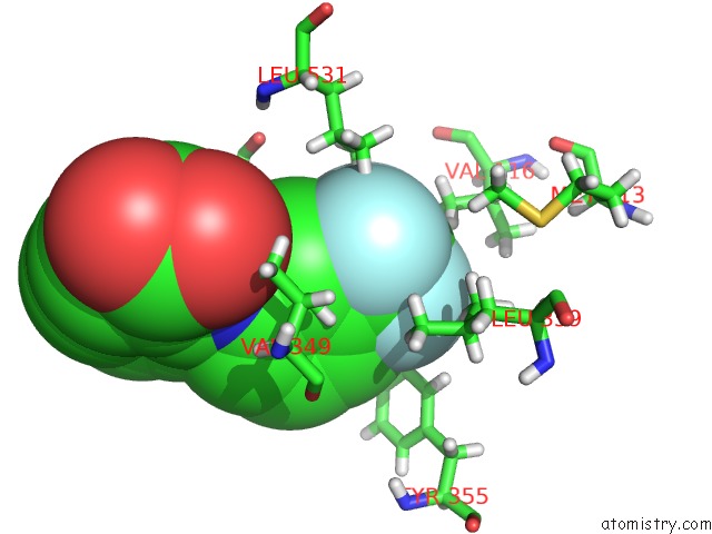
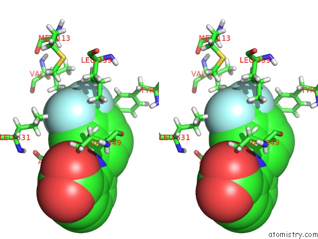
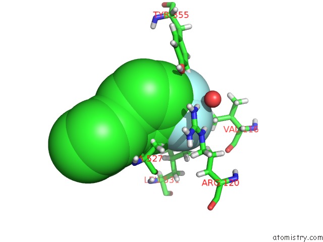
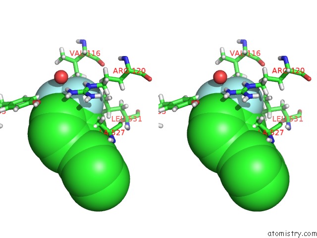
Fluorine binding site 1 out of 6 in 5ikv
Go back to
Fluorine binding site 1 out
of 6 in the The Structure of Flufenamic Acid Bound to Human Cyclooxygenase-2
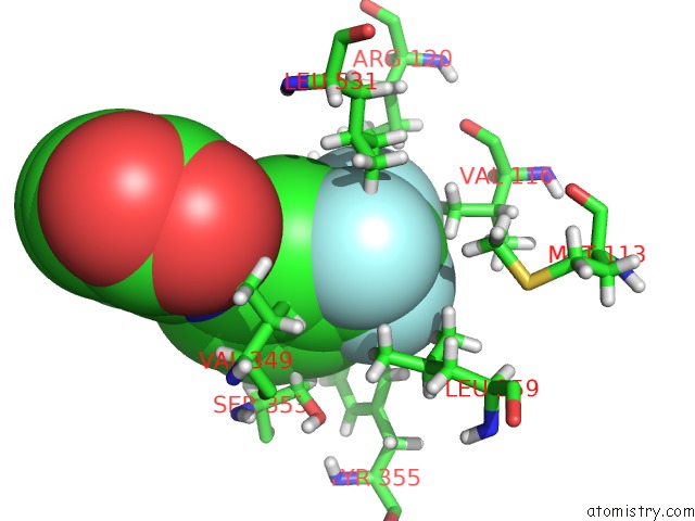
Mono view
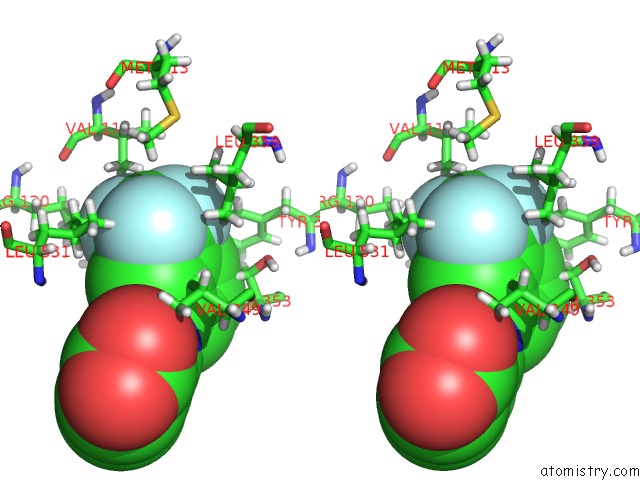
Stereo pair view
Mono view
Stereo pair view
A full contact list of Fluorine with other atoms in the F binding
site number 1 of The Structure of Flufenamic Acid Bound to Human Cyclooxygenase-2 within 5.0Å range:
|
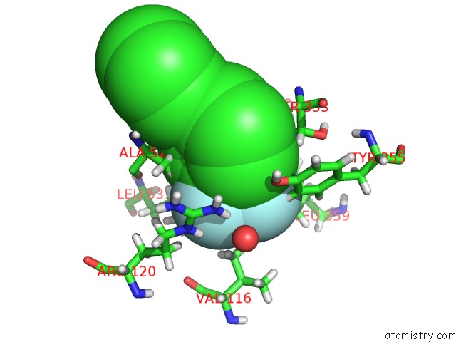
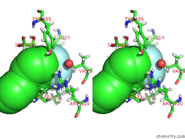
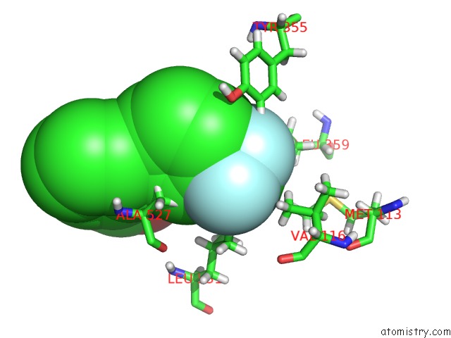
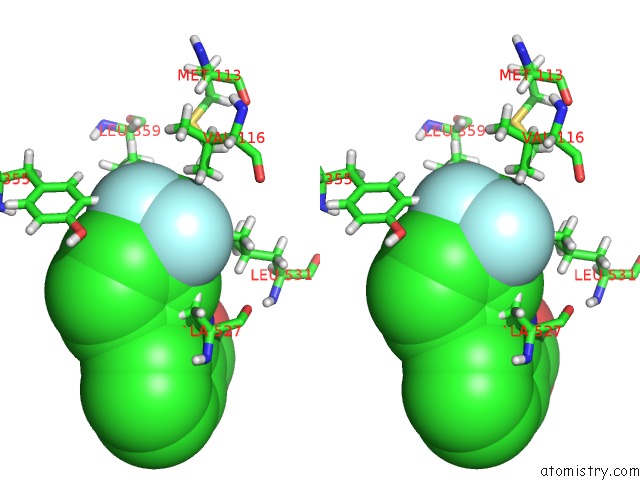
Fluorine binding site 2 out of 6 in 5ikv
Go back to
Fluorine binding site 2 out
of 6 in the The Structure of Flufenamic Acid Bound to Human Cyclooxygenase-2
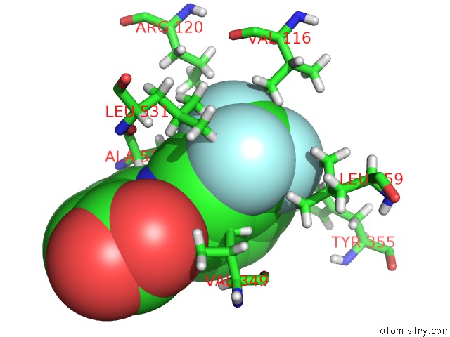
Mono view
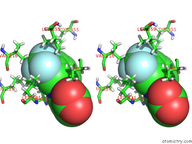
Stereo pair view
Mono view
Stereo pair view
A full contact list of Fluorine with other atoms in the F binding
site number 2 of The Structure of Flufenamic Acid Bound to Human Cyclooxygenase-2 within 5.0Å range:
|
Fluorine binding site 3 out of 6 in 5ikv
Go back to
Fluorine binding site 3 out
of 6 in the The Structure of Flufenamic Acid Bound to Human Cyclooxygenase-2
Mono view
Stereo pair view
Mono view
Stereo pair view
A full contact list of Fluorine with other atoms in the F binding
site number 3 of The Structure of Flufenamic Acid Bound to Human Cyclooxygenase-2 within 5.0Å range:
|
Fluorine binding site 4 out of 6 in 5ikv
Go back to
Fluorine binding site 4 out
of 6 in the The Structure of Flufenamic Acid Bound to Human Cyclooxygenase-2
Mono view
Stereo pair view
Mono view
Stereo pair view
A full contact list of Fluorine with other atoms in the F binding
site number 4 of The Structure of Flufenamic Acid Bound to Human Cyclooxygenase-2 within 5.0Å range:
|
Fluorine binding site 5 out of 6 in 5ikv
Go back to
Fluorine binding site 5 out
of 6 in the The Structure of Flufenamic Acid Bound to Human Cyclooxygenase-2
Mono view
Stereo pair view
Mono view
Stereo pair view
A full contact list of Fluorine with other atoms in the F binding
site number 5 of The Structure of Flufenamic Acid Bound to Human Cyclooxygenase-2 within 5.0Å range:
|
Fluorine binding site 6 out of 6 in 5ikv
Go back to
Fluorine binding site 6 out
of 6 in the The Structure of Flufenamic Acid Bound to Human Cyclooxygenase-2
Mono view
Stereo pair view
Mono view
Stereo pair view
A full contact list of Fluorine with other atoms in the F binding
site number 6 of The Structure of Flufenamic Acid Bound to Human Cyclooxygenase-2 within 5.0Å range:
|
Reference:
B.J.Orlando,
M.G.Malkowski.
Substrate-Selective Inhibition of Cyclooxygeanse-2 By Fenamic Acid Derivatives Is Dependent on Peroxide Tone. J.Biol.Chem. V. 291 15069 2016.
ISSN: ESSN 1083-351X
PubMed: 27226593
DOI: 10.1074/JBC.M116.725713
Page generated: Tue Jul 15 04:09:36 2025
ISSN: ESSN 1083-351X
PubMed: 27226593
DOI: 10.1074/JBC.M116.725713
Last articles
K in 7F1AK in 7F0N
K in 7EIA
K in 7DFY
K in 7EAY
K in 7E20
K in 7DVQ
K in 7D5E
K in 7DMR
K in 7DIF