Fluorine »
PDB 5lz7-5msb »
5mmb »
Fluorine in PDB 5mmb: Crystal Structure of the Prototype Foamy Virus (Pfv) Intasome in Complex with Magnesium and the Insti XZ434 (Compound 6P)
Enzymatic activity of Crystal Structure of the Prototype Foamy Virus (Pfv) Intasome in Complex with Magnesium and the Insti XZ434 (Compound 6P)
All present enzymatic activity of Crystal Structure of the Prototype Foamy Virus (Pfv) Intasome in Complex with Magnesium and the Insti XZ434 (Compound 6P):
2.7.7.49; 2.7.7.7; 3.1.26.4;
2.7.7.49; 2.7.7.7; 3.1.26.4;
Protein crystallography data
The structure of Crystal Structure of the Prototype Foamy Virus (Pfv) Intasome in Complex with Magnesium and the Insti XZ434 (Compound 6P), PDB code: 5mmb
was solved by
D.P.Maskell,
V.E.Pye,
P.Cherepanov,
with X-Ray Crystallography technique. A brief refinement statistics is given in the table below:
| Resolution Low / High (Å) | 57.77 / 2.77 |
| Space group | P 41 21 2 |
| Cell size a, b, c (Å), α, β, γ (°) | 160.490, 160.490, 123.830, 90.00, 90.00, 90.00 |
| R / Rfree (%) | 18.4 / 20.3 |
Other elements in 5mmb:
The structure of Crystal Structure of the Prototype Foamy Virus (Pfv) Intasome in Complex with Magnesium and the Insti XZ434 (Compound 6P) also contains other interesting chemical elements:
| Magnesium | (Mg) | 3 atoms |
| Zinc | (Zn) | 1 atom |
Fluorine Binding Sites:
The binding sites of Fluorine atom in the Crystal Structure of the Prototype Foamy Virus (Pfv) Intasome in Complex with Magnesium and the Insti XZ434 (Compound 6P)
(pdb code 5mmb). This binding sites where shown within
5.0 Angstroms radius around Fluorine atom.
In total 2 binding sites of Fluorine where determined in the Crystal Structure of the Prototype Foamy Virus (Pfv) Intasome in Complex with Magnesium and the Insti XZ434 (Compound 6P), PDB code: 5mmb:
Jump to Fluorine binding site number: 1; 2;
In total 2 binding sites of Fluorine where determined in the Crystal Structure of the Prototype Foamy Virus (Pfv) Intasome in Complex with Magnesium and the Insti XZ434 (Compound 6P), PDB code: 5mmb:
Jump to Fluorine binding site number: 1; 2;
Fluorine binding site 1 out of 2 in 5mmb
Go back to
Fluorine binding site 1 out
of 2 in the Crystal Structure of the Prototype Foamy Virus (Pfv) Intasome in Complex with Magnesium and the Insti XZ434 (Compound 6P)
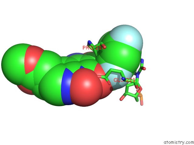
Mono view
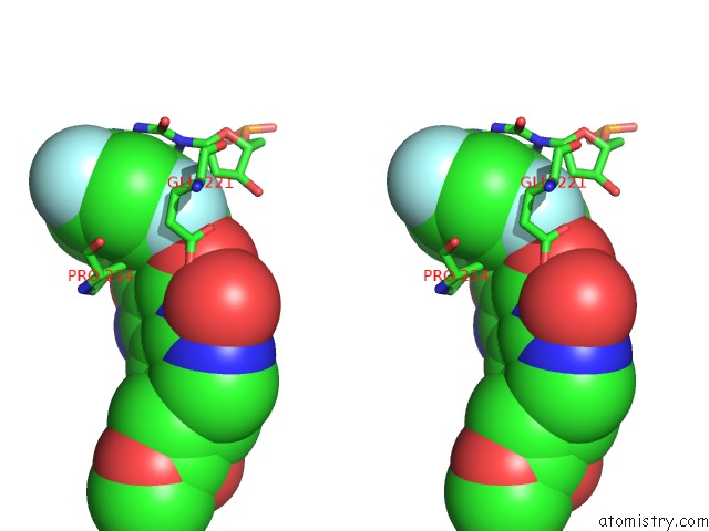
Stereo pair view
Mono view
Stereo pair view
A full contact list of Fluorine with other atoms in the F binding
site number 1 of Crystal Structure of the Prototype Foamy Virus (Pfv) Intasome in Complex with Magnesium and the Insti XZ434 (Compound 6P) within 5.0Å range:
|
Fluorine binding site 2 out of 2 in 5mmb
Go back to
Fluorine binding site 2 out
of 2 in the Crystal Structure of the Prototype Foamy Virus (Pfv) Intasome in Complex with Magnesium and the Insti XZ434 (Compound 6P)
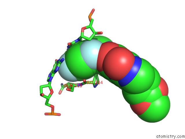
Mono view
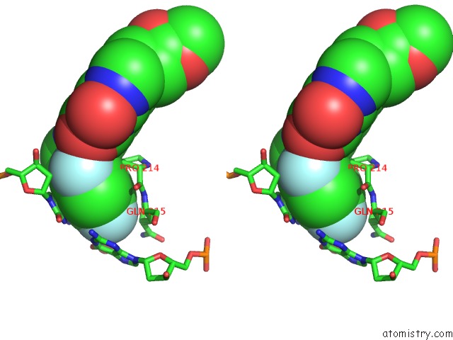
Stereo pair view
Mono view
Stereo pair view
A full contact list of Fluorine with other atoms in the F binding
site number 2 of Crystal Structure of the Prototype Foamy Virus (Pfv) Intasome in Complex with Magnesium and the Insti XZ434 (Compound 6P) within 5.0Å range:
|
Reference:
X.Z.Zhao,
S.J.Smith,
D.P.Maskell,
M.Metifiot,
V.E.Pye,
K.Fesen,
C.Marchand,
Y.Pommier,
P.Cherepanov,
S.H.Hughes,
T.R.Burke.
Structure-Guided Optimization of Hiv Integrase Strand Transfer Inhibitors. J. Med. Chem. V. 60 7315 2017.
ISSN: ISSN 1520-4804
PubMed: 28737946
DOI: 10.1021/ACS.JMEDCHEM.7B00596
Page generated: Tue Jul 15 05:16:23 2025
ISSN: ISSN 1520-4804
PubMed: 28737946
DOI: 10.1021/ACS.JMEDCHEM.7B00596
Last articles
Mg in 4DV2Mg in 4DV0
Mg in 4DV1
Mg in 4DUZ
Mg in 4DUY
Mg in 4DR7
Mg in 4DR6
Mg in 4DR5
Mg in 4DUX
Mg in 4DUW