Fluorine »
PDB 5nk4-5o6j »
5nx2 »
Fluorine in PDB 5nx2: Crystal Structure of Thermostabilised Full-Length Glp-1R in Complex with A Truncated Peptide Agonist at 3.7 A Resolution
Protein crystallography data
The structure of Crystal Structure of Thermostabilised Full-Length Glp-1R in Complex with A Truncated Peptide Agonist at 3.7 A Resolution, PDB code: 5nx2
was solved by
M.Rappas,
A.Jazayeri,
A.J.H.Brown,
J.Kean,
J.C.Errey,
N.Robertson,
C.Fiez-Vandal,
S.P.Andrews,
M.Congreve,
A.Bortolato,
J.S.Mason,
A.H.Baig,
I.Teobald,
A.S.Dore,
M.Weir,
R.M.Cooke,
F.H.Marshall,
with X-Ray Crystallography technique. A brief refinement statistics is given in the table below:
| Resolution Low / High (Å) | 24.68 / 3.70 |
| Space group | P 31 2 1 |
| Cell size a, b, c (Å), α, β, γ (°) | 94.435, 94.435, 163.905, 90.00, 90.00, 120.00 |
| R / Rfree (%) | 28.5 / 33.4 |
Fluorine Binding Sites:
The binding sites of Fluorine atom in the Crystal Structure of Thermostabilised Full-Length Glp-1R in Complex with A Truncated Peptide Agonist at 3.7 A Resolution
(pdb code 5nx2). This binding sites where shown within
5.0 Angstroms radius around Fluorine atom.
In total only one binding site of Fluorine was determined in the Crystal Structure of Thermostabilised Full-Length Glp-1R in Complex with A Truncated Peptide Agonist at 3.7 A Resolution, PDB code: 5nx2:
In total only one binding site of Fluorine was determined in the Crystal Structure of Thermostabilised Full-Length Glp-1R in Complex with A Truncated Peptide Agonist at 3.7 A Resolution, PDB code: 5nx2:
Fluorine binding site 1 out of 1 in 5nx2
Go back to
Fluorine binding site 1 out
of 1 in the Crystal Structure of Thermostabilised Full-Length Glp-1R in Complex with A Truncated Peptide Agonist at 3.7 A Resolution
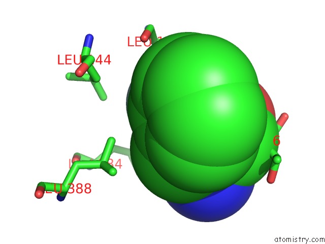
Mono view
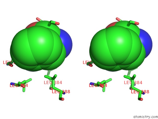
Stereo pair view
Mono view
Stereo pair view
A full contact list of Fluorine with other atoms in the F binding
site number 1 of Crystal Structure of Thermostabilised Full-Length Glp-1R in Complex with A Truncated Peptide Agonist at 3.7 A Resolution within 5.0Å range:
|
Reference:
A.Jazayeri,
M.Rappas,
A.J.H.Brown,
J.Kean,
J.C.Errey,
N.J.Robertson,
C.Fiez-Vandal,
S.P.Andrews,
M.Congreve,
A.Bortolato,
J.S.Mason,
A.H.Baig,
I.Teobald,
A.S.Dore,
M.Weir,
R.M.Cooke,
F.H.Marshall.
Crystal Structure of the Glp-1 Receptor Bound to A Peptide Agonist. Nature V. 546 254 2017.
ISSN: ISSN 0028-0836
PubMed: 28562585
DOI: 10.1038/NATURE22800
Page generated: Tue Jul 15 05:34:35 2025
ISSN: ISSN 0028-0836
PubMed: 28562585
DOI: 10.1038/NATURE22800
Last articles
Na in 5YDBNa in 5YID
Na in 5YBZ
Na in 5YDD
Na in 5Y7D
Na in 5YAO
Na in 5YAX
Na in 5YAL
Na in 5Y94
Na in 5Y3E