Fluorine »
PDB 5olx-5p93 »
5ouj »
Fluorine in PDB 5ouj: Crystal Structure of Human AKR1B1 Complexed with Nadp+ and Compound 39
Enzymatic activity of Crystal Structure of Human AKR1B1 Complexed with Nadp+ and Compound 39
All present enzymatic activity of Crystal Structure of Human AKR1B1 Complexed with Nadp+ and Compound 39:
1.1.1.21;
1.1.1.21;
Protein crystallography data
The structure of Crystal Structure of Human AKR1B1 Complexed with Nadp+ and Compound 39, PDB code: 5ouj
was solved by
A.Cousido-Siah,
F.X.Ruiz,
A.Mitschler,
K.Metwally,
A.Podjarny,
with X-Ray Crystallography technique. A brief refinement statistics is given in the table below:
| Resolution Low / High (Å) | 28.90 / 0.96 |
| Space group | P 1 |
| Cell size a, b, c (Å), α, β, γ (°) | 40.050, 46.979, 47.283, 76.40, 67.57, 77.40 |
| R / Rfree (%) | 13 / 14.7 |
Other elements in 5ouj:
The structure of Crystal Structure of Human AKR1B1 Complexed with Nadp+ and Compound 39 also contains other interesting chemical elements:
| Chlorine | (Cl) | 1 atom |
Fluorine Binding Sites:
The binding sites of Fluorine atom in the Crystal Structure of Human AKR1B1 Complexed with Nadp+ and Compound 39
(pdb code 5ouj). This binding sites where shown within
5.0 Angstroms radius around Fluorine atom.
In total only one binding site of Fluorine was determined in the Crystal Structure of Human AKR1B1 Complexed with Nadp+ and Compound 39, PDB code: 5ouj:
In total only one binding site of Fluorine was determined in the Crystal Structure of Human AKR1B1 Complexed with Nadp+ and Compound 39, PDB code: 5ouj:
Fluorine binding site 1 out of 1 in 5ouj
Go back to
Fluorine binding site 1 out
of 1 in the Crystal Structure of Human AKR1B1 Complexed with Nadp+ and Compound 39
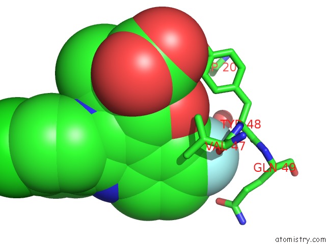
Mono view
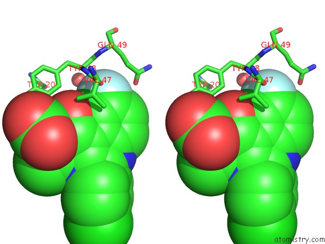
Stereo pair view
Mono view
Stereo pair view
A full contact list of Fluorine with other atoms in the F binding
site number 1 of Crystal Structure of Human AKR1B1 Complexed with Nadp+ and Compound 39 within 5.0Å range:
|
Reference:
I.Crespo,
J.Gimenez-Dejoz,
S.Porte,
A.Cousido-Siah,
A.Mitschler,
A.Podjarny,
H.Pratsinis,
D.Kletsas,
X.Pares,
F.X.Ruiz,
K.Metwally,
J.Farres.
Design, Synthesis, Structure-Activity Relationships and X-Ray Structural Studies of Novel 1-Oxopyrimido[4,5-C]Quinoline-2-Acetic Acid Derivatives As Selective and Potent Inhibitors of Human Aldose Reductase. Eur J Med Chem V. 152 160 2018.
ISSN: ISSN 1768-3254
PubMed: 29705708
DOI: 10.1016/J.EJMECH.2018.04.015
Page generated: Tue Jul 15 05:55:53 2025
ISSN: ISSN 1768-3254
PubMed: 29705708
DOI: 10.1016/J.EJMECH.2018.04.015
Last articles
Mg in 6DW5Mg in 6DW7
Mg in 6DW4
Mg in 6DW3
Mg in 6DUQ
Mg in 6DVK
Mg in 6DUK
Mg in 6DV9
Mg in 6DUS
Mg in 6DUH