Fluorine »
PDB 5sb4-5sjr »
5sem »
Fluorine in PDB 5sem: Crystal Structure of Human Phosphodiesterase 10 in Complex with 3- Methyl-6-[2-(1-Methyl-4-Phenylimidazol-2-Yl)Ethyl]-2- (Trifluoromethyl)Imidazo[1,2-B]Pyridazine
Enzymatic activity of Crystal Structure of Human Phosphodiesterase 10 in Complex with 3- Methyl-6-[2-(1-Methyl-4-Phenylimidazol-2-Yl)Ethyl]-2- (Trifluoromethyl)Imidazo[1,2-B]Pyridazine
All present enzymatic activity of Crystal Structure of Human Phosphodiesterase 10 in Complex with 3- Methyl-6-[2-(1-Methyl-4-Phenylimidazol-2-Yl)Ethyl]-2- (Trifluoromethyl)Imidazo[1,2-B]Pyridazine:
3.1.4.17;
3.1.4.17;
Protein crystallography data
The structure of Crystal Structure of Human Phosphodiesterase 10 in Complex with 3- Methyl-6-[2-(1-Methyl-4-Phenylimidazol-2-Yl)Ethyl]-2- (Trifluoromethyl)Imidazo[1,2-B]Pyridazine, PDB code: 5sem
was solved by
C.Joseph,
K.Groebke-Zbinden,
J.Benz,
D.Schlatter,
M.G.Rudolph,
with X-Ray Crystallography technique. A brief refinement statistics is given in the table below:
| Resolution Low / High (Å) | 43.57 / 2.10 |
| Space group | H 3 |
| Cell size a, b, c (Å), α, β, γ (°) | 135.449, 135.449, 235.505, 90, 90, 120 |
| R / Rfree (%) | 17.8 / 22.8 |
Other elements in 5sem:
The structure of Crystal Structure of Human Phosphodiesterase 10 in Complex with 3- Methyl-6-[2-(1-Methyl-4-Phenylimidazol-2-Yl)Ethyl]-2- (Trifluoromethyl)Imidazo[1,2-B]Pyridazine also contains other interesting chemical elements:
| Magnesium | (Mg) | 4 atoms |
| Zinc | (Zn) | 4 atoms |
Fluorine Binding Sites:
Pages:
>>> Page 1 <<< Page 2, Binding sites: 11 - 12;Binding sites:
The binding sites of Fluorine atom in the Crystal Structure of Human Phosphodiesterase 10 in Complex with 3- Methyl-6-[2-(1-Methyl-4-Phenylimidazol-2-Yl)Ethyl]-2- (Trifluoromethyl)Imidazo[1,2-B]Pyridazine (pdb code 5sem). This binding sites where shown within 5.0 Angstroms radius around Fluorine atom.In total 12 binding sites of Fluorine where determined in the Crystal Structure of Human Phosphodiesterase 10 in Complex with 3- Methyl-6-[2-(1-Methyl-4-Phenylimidazol-2-Yl)Ethyl]-2- (Trifluoromethyl)Imidazo[1,2-B]Pyridazine, PDB code: 5sem:
Jump to Fluorine binding site number: 1; 2; 3; 4; 5; 6; 7; 8; 9; 10;
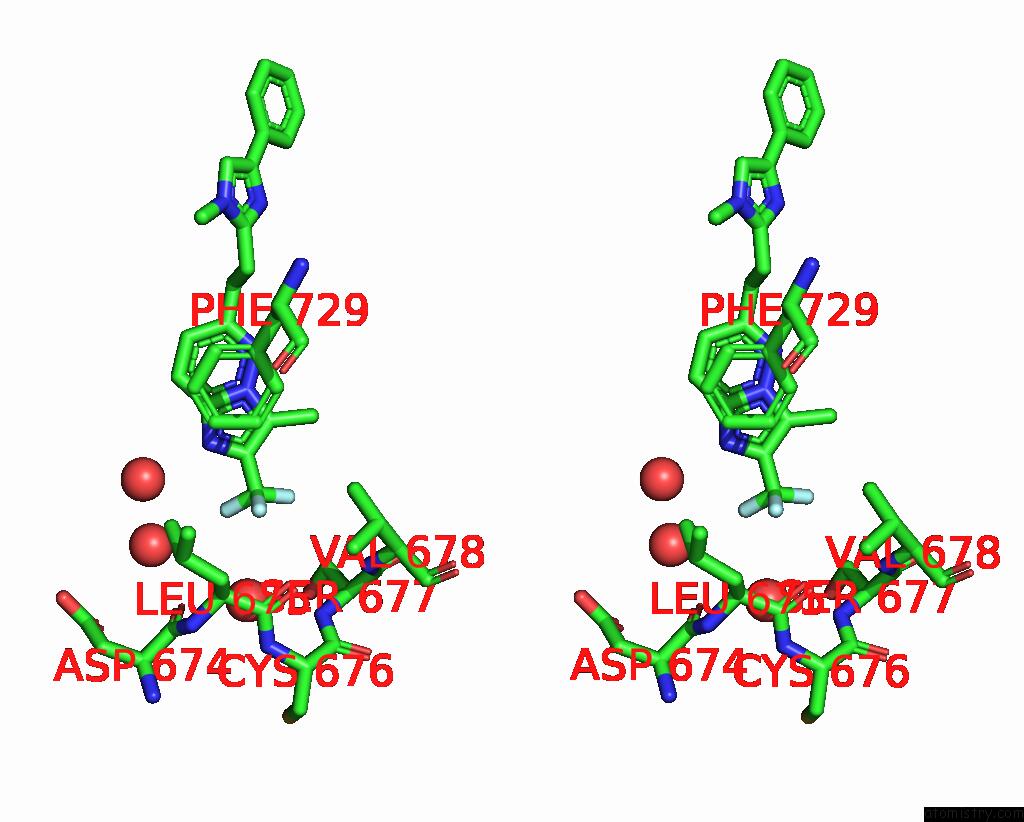
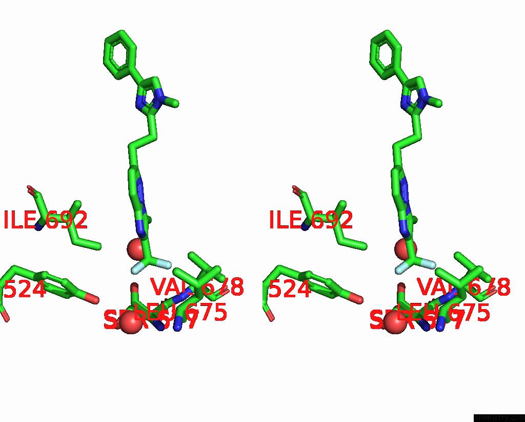
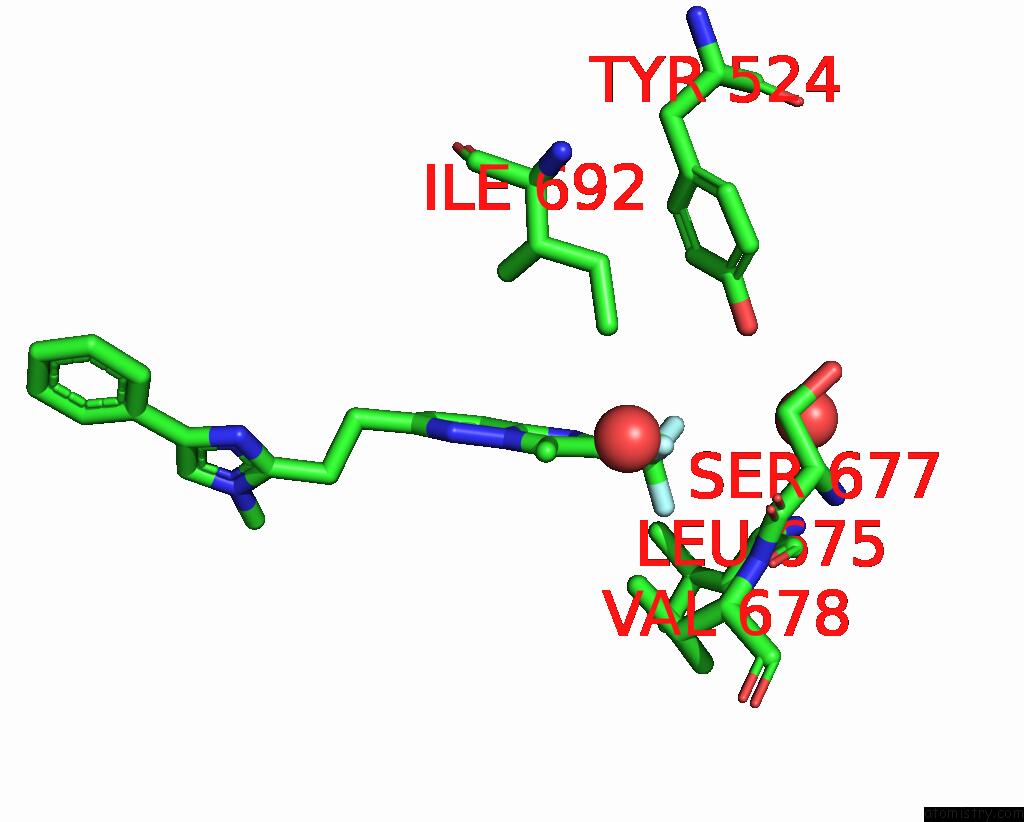
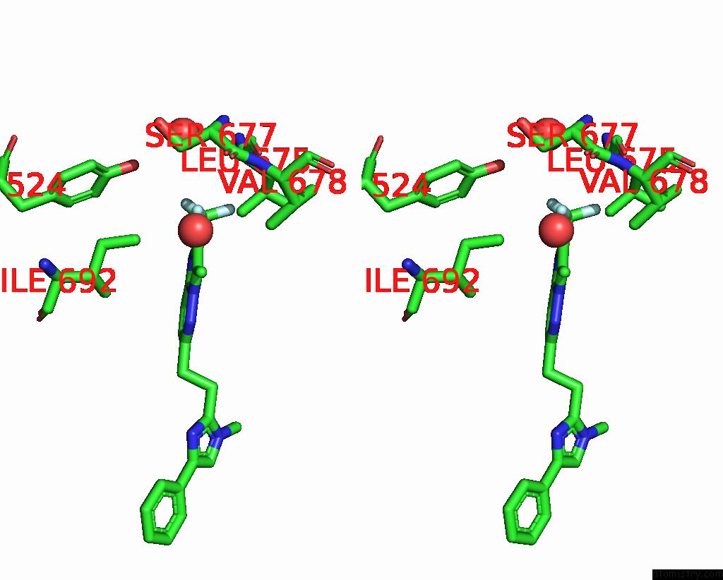
Fluorine binding site 1 out of 12 in 5sem
Go back to
Fluorine binding site 1 out
of 12 in the Crystal Structure of Human Phosphodiesterase 10 in Complex with 3- Methyl-6-[2-(1-Methyl-4-Phenylimidazol-2-Yl)Ethyl]-2- (Trifluoromethyl)Imidazo[1,2-B]Pyridazine
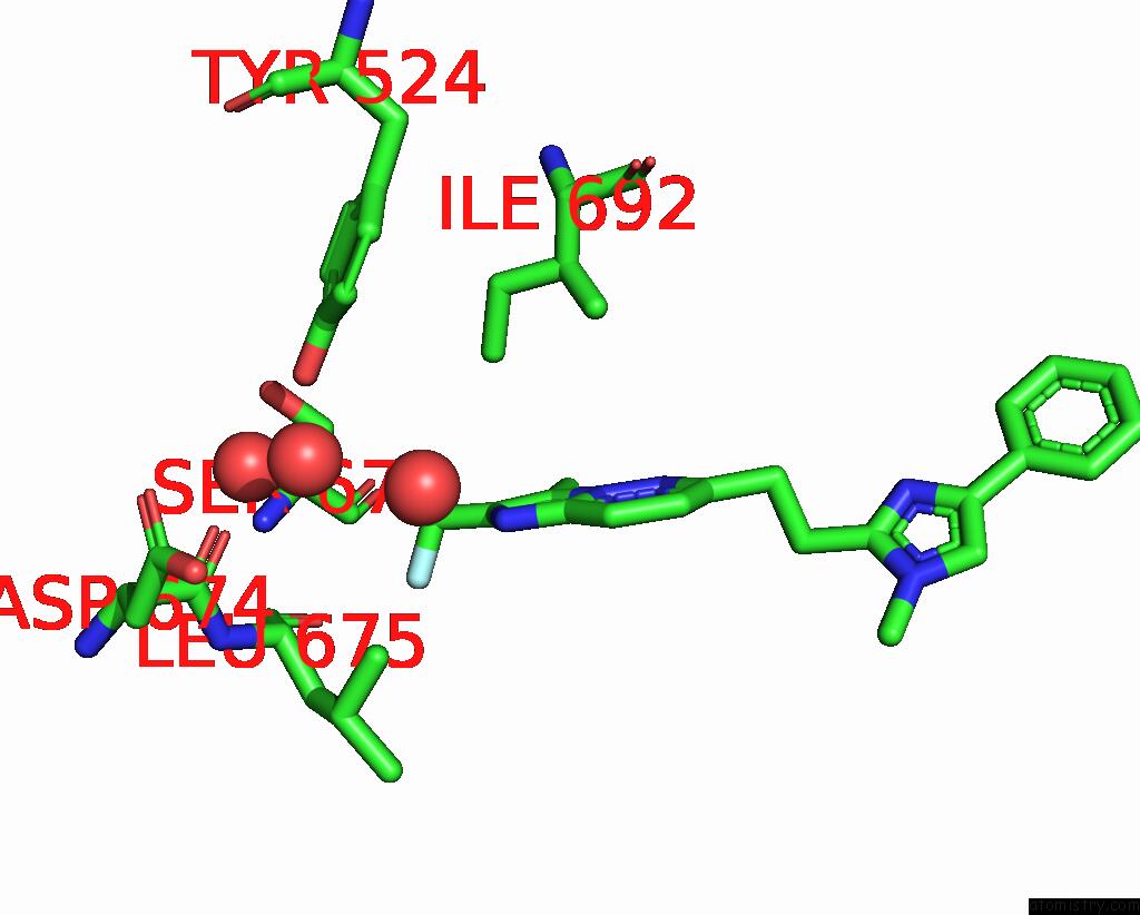
Mono view
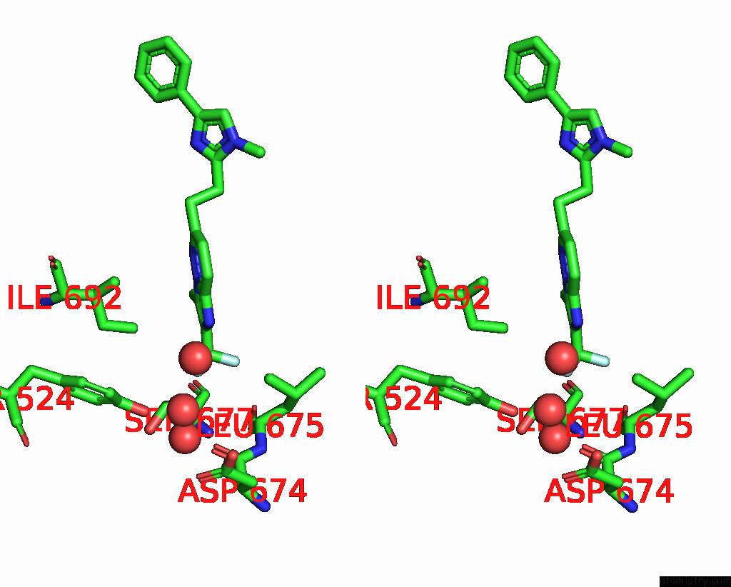
Stereo pair view
Mono view
Stereo pair view
A full contact list of Fluorine with other atoms in the F binding
site number 1 of Crystal Structure of Human Phosphodiesterase 10 in Complex with 3- Methyl-6-[2-(1-Methyl-4-Phenylimidazol-2-Yl)Ethyl]-2- (Trifluoromethyl)Imidazo[1,2-B]Pyridazine within 5.0Å range:
|
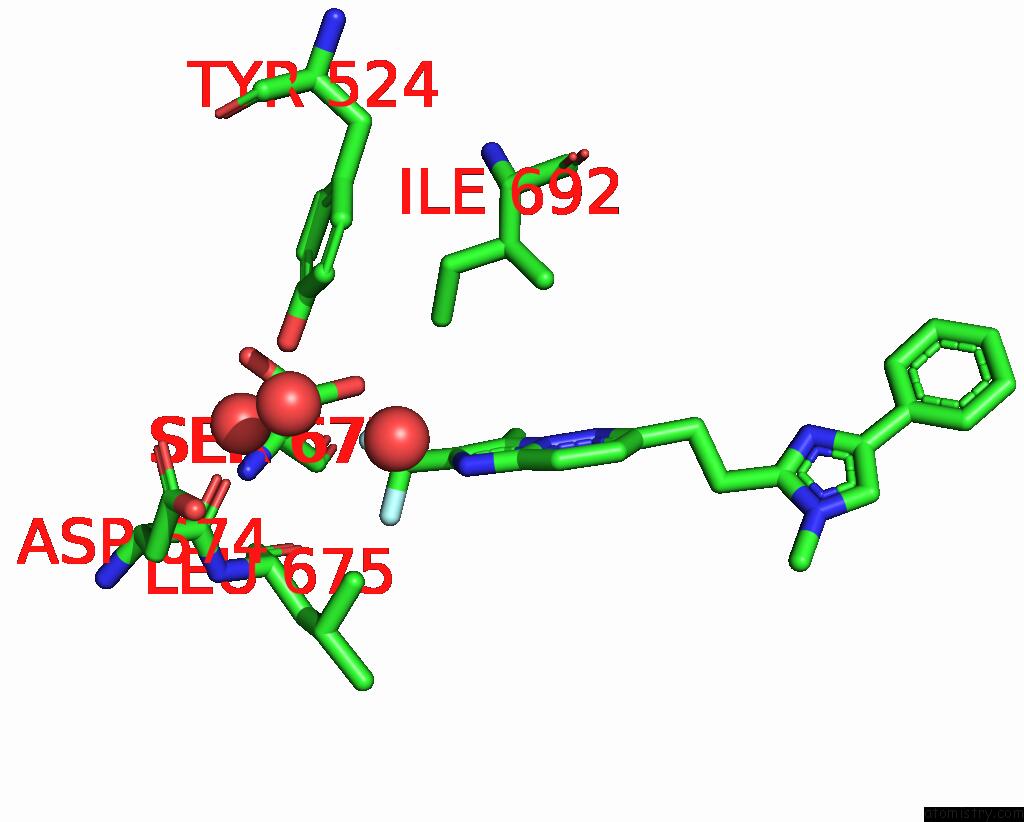
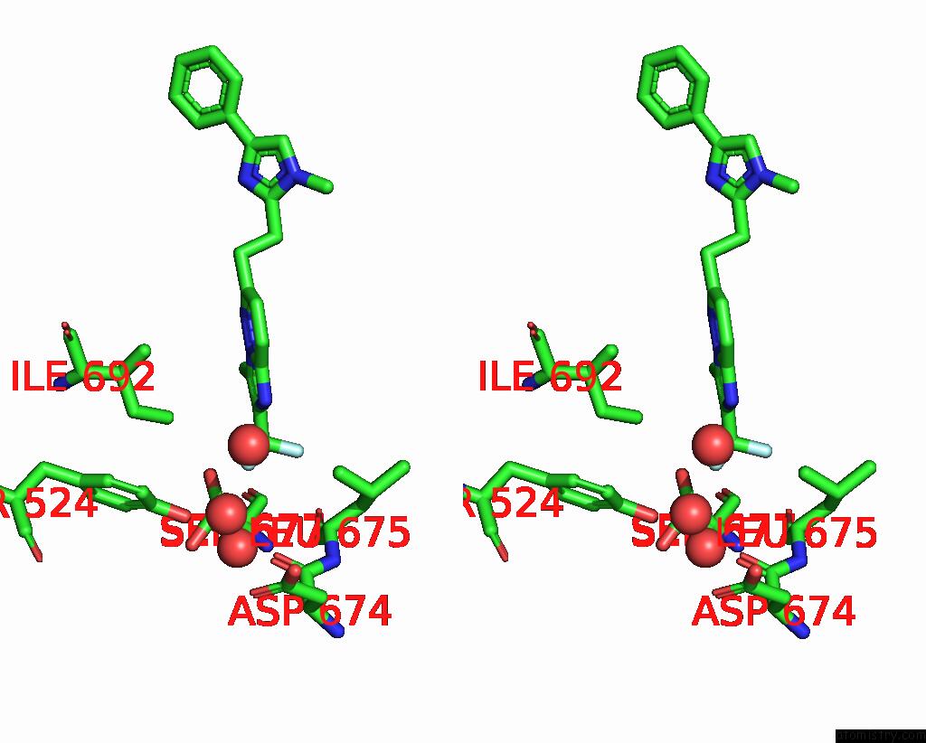
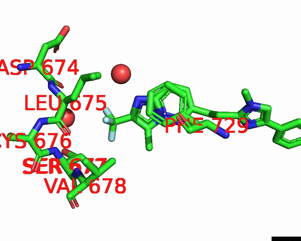
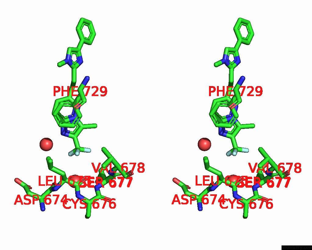
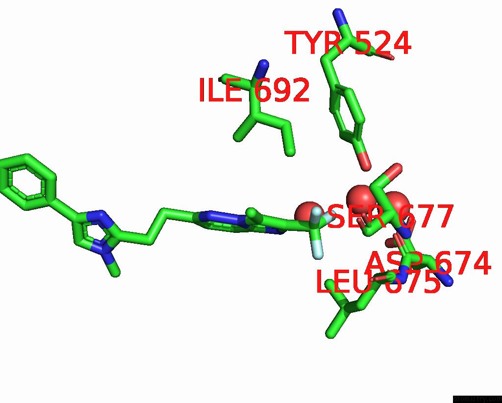
Fluorine binding site 2 out of 12 in 5sem
Go back to
Fluorine binding site 2 out
of 12 in the Crystal Structure of Human Phosphodiesterase 10 in Complex with 3- Methyl-6-[2-(1-Methyl-4-Phenylimidazol-2-Yl)Ethyl]-2- (Trifluoromethyl)Imidazo[1,2-B]Pyridazine
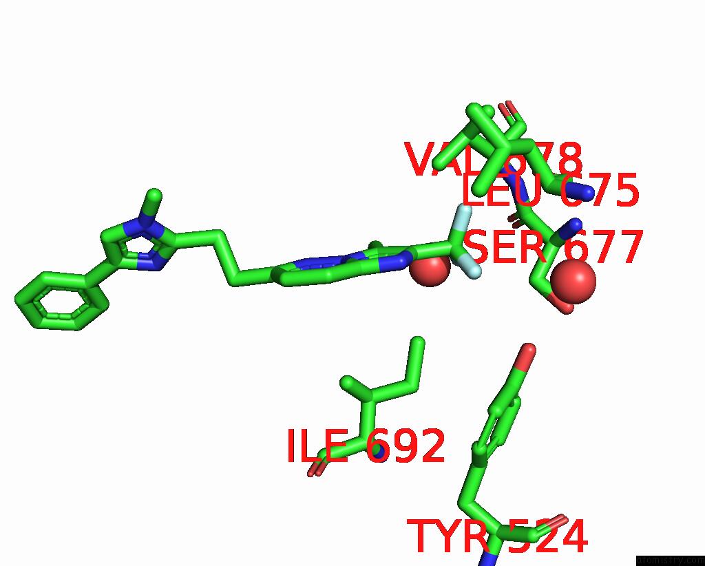
Mono view
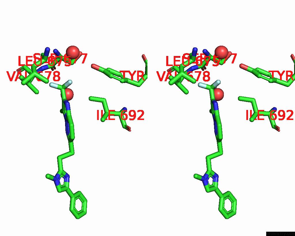
Stereo pair view
Mono view
Stereo pair view
A full contact list of Fluorine with other atoms in the F binding
site number 2 of Crystal Structure of Human Phosphodiesterase 10 in Complex with 3- Methyl-6-[2-(1-Methyl-4-Phenylimidazol-2-Yl)Ethyl]-2- (Trifluoromethyl)Imidazo[1,2-B]Pyridazine within 5.0Å range:
|
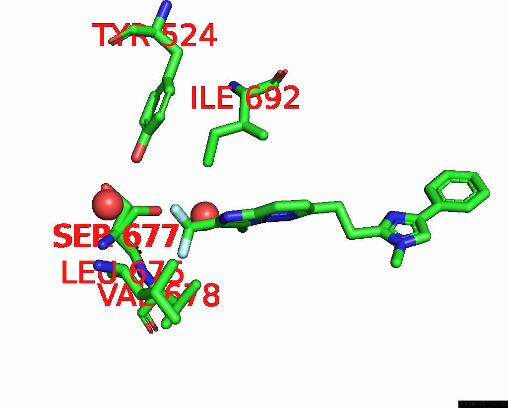
Fluorine binding site 3 out of 12 in 5sem
Go back to
Fluorine binding site 3 out
of 12 in the Crystal Structure of Human Phosphodiesterase 10 in Complex with 3- Methyl-6-[2-(1-Methyl-4-Phenylimidazol-2-Yl)Ethyl]-2- (Trifluoromethyl)Imidazo[1,2-B]Pyridazine

Mono view
Stereo pair view
Mono view
Stereo pair view
A full contact list of Fluorine with other atoms in the F binding
site number 3 of Crystal Structure of Human Phosphodiesterase 10 in Complex with 3- Methyl-6-[2-(1-Methyl-4-Phenylimidazol-2-Yl)Ethyl]-2- (Trifluoromethyl)Imidazo[1,2-B]Pyridazine within 5.0Å range:
|
Fluorine binding site 4 out of 12 in 5sem
Go back to
Fluorine binding site 4 out
of 12 in the Crystal Structure of Human Phosphodiesterase 10 in Complex with 3- Methyl-6-[2-(1-Methyl-4-Phenylimidazol-2-Yl)Ethyl]-2- (Trifluoromethyl)Imidazo[1,2-B]Pyridazine
Mono view
Stereo pair view
Mono view
Stereo pair view
A full contact list of Fluorine with other atoms in the F binding
site number 4 of Crystal Structure of Human Phosphodiesterase 10 in Complex with 3- Methyl-6-[2-(1-Methyl-4-Phenylimidazol-2-Yl)Ethyl]-2- (Trifluoromethyl)Imidazo[1,2-B]Pyridazine within 5.0Å range:
|
Fluorine binding site 5 out of 12 in 5sem
Go back to
Fluorine binding site 5 out
of 12 in the Crystal Structure of Human Phosphodiesterase 10 in Complex with 3- Methyl-6-[2-(1-Methyl-4-Phenylimidazol-2-Yl)Ethyl]-2- (Trifluoromethyl)Imidazo[1,2-B]Pyridazine
Mono view
Stereo pair view
Mono view
Stereo pair view
A full contact list of Fluorine with other atoms in the F binding
site number 5 of Crystal Structure of Human Phosphodiesterase 10 in Complex with 3- Methyl-6-[2-(1-Methyl-4-Phenylimidazol-2-Yl)Ethyl]-2- (Trifluoromethyl)Imidazo[1,2-B]Pyridazine within 5.0Å range:
|
Fluorine binding site 6 out of 12 in 5sem
Go back to
Fluorine binding site 6 out
of 12 in the Crystal Structure of Human Phosphodiesterase 10 in Complex with 3- Methyl-6-[2-(1-Methyl-4-Phenylimidazol-2-Yl)Ethyl]-2- (Trifluoromethyl)Imidazo[1,2-B]Pyridazine
Mono view
Stereo pair view
Mono view
Stereo pair view
A full contact list of Fluorine with other atoms in the F binding
site number 6 of Crystal Structure of Human Phosphodiesterase 10 in Complex with 3- Methyl-6-[2-(1-Methyl-4-Phenylimidazol-2-Yl)Ethyl]-2- (Trifluoromethyl)Imidazo[1,2-B]Pyridazine within 5.0Å range:
|
Fluorine binding site 7 out of 12 in 5sem
Go back to
Fluorine binding site 7 out
of 12 in the Crystal Structure of Human Phosphodiesterase 10 in Complex with 3- Methyl-6-[2-(1-Methyl-4-Phenylimidazol-2-Yl)Ethyl]-2- (Trifluoromethyl)Imidazo[1,2-B]Pyridazine

Mono view

Stereo pair view
Mono view
Stereo pair view
A full contact list of Fluorine with other atoms in the F binding
site number 7 of Crystal Structure of Human Phosphodiesterase 10 in Complex with 3- Methyl-6-[2-(1-Methyl-4-Phenylimidazol-2-Yl)Ethyl]-2- (Trifluoromethyl)Imidazo[1,2-B]Pyridazine within 5.0Å range:
|
Fluorine binding site 8 out of 12 in 5sem
Go back to
Fluorine binding site 8 out
of 12 in the Crystal Structure of Human Phosphodiesterase 10 in Complex with 3- Methyl-6-[2-(1-Methyl-4-Phenylimidazol-2-Yl)Ethyl]-2- (Trifluoromethyl)Imidazo[1,2-B]Pyridazine
Mono view
Stereo pair view
Mono view
Stereo pair view
A full contact list of Fluorine with other atoms in the F binding
site number 8 of Crystal Structure of Human Phosphodiesterase 10 in Complex with 3- Methyl-6-[2-(1-Methyl-4-Phenylimidazol-2-Yl)Ethyl]-2- (Trifluoromethyl)Imidazo[1,2-B]Pyridazine within 5.0Å range:
|
Fluorine binding site 9 out of 12 in 5sem
Go back to
Fluorine binding site 9 out
of 12 in the Crystal Structure of Human Phosphodiesterase 10 in Complex with 3- Methyl-6-[2-(1-Methyl-4-Phenylimidazol-2-Yl)Ethyl]-2- (Trifluoromethyl)Imidazo[1,2-B]Pyridazine

Mono view

Stereo pair view
Mono view
Stereo pair view
A full contact list of Fluorine with other atoms in the F binding
site number 9 of Crystal Structure of Human Phosphodiesterase 10 in Complex with 3- Methyl-6-[2-(1-Methyl-4-Phenylimidazol-2-Yl)Ethyl]-2- (Trifluoromethyl)Imidazo[1,2-B]Pyridazine within 5.0Å range:
|
Fluorine binding site 10 out of 12 in 5sem
Go back to
Fluorine binding site 10 out
of 12 in the Crystal Structure of Human Phosphodiesterase 10 in Complex with 3- Methyl-6-[2-(1-Methyl-4-Phenylimidazol-2-Yl)Ethyl]-2- (Trifluoromethyl)Imidazo[1,2-B]Pyridazine
Mono view

Stereo pair view
Mono view
Stereo pair view
A full contact list of Fluorine with other atoms in the F binding
site number 10 of Crystal Structure of Human Phosphodiesterase 10 in Complex with 3- Methyl-6-[2-(1-Methyl-4-Phenylimidazol-2-Yl)Ethyl]-2- (Trifluoromethyl)Imidazo[1,2-B]Pyridazine within 5.0Å range:
|
Reference:
A.Tosstorff,
M.G.Rudolph,
J.C.Cole,
M.Reutlinger,
C.Kramer,
H.Schaffhauser,
A.Nilly,
A.Flohr,
B.Kuhn.
A High Quality, Industrial Data Set For Binding Affinity Prediction: Performance Comparison in Different Early Drug Discovery Scenarios. J.Comput.Aided Mol.Des. V. 36 753 2022.
ISSN: ESSN 1573-4951
PubMed: 36153472
DOI: 10.1007/S10822-022-00478-X
Page generated: Tue Jul 15 07:09:45 2025
ISSN: ESSN 1573-4951
PubMed: 36153472
DOI: 10.1007/S10822-022-00478-X
Last articles
Mn in 2D3RMn in 2D3P
Mn in 2CEV
Mn in 2CV5
Mn in 2CYF
Mn in 2D0C
Mn in 2CY6
Mn in 2CWM
Mn in 2CTV
Mn in 2CM1