Fluorine »
PDB 5sk2-5soj »
5so3 »
Fluorine in PDB 5so3: Pandda Analysis Group Deposition -- Crystal Structure of Pseudomonas Aeruginosa Fabf-C164Q Mutant Protein in Complex with JKH100B
Enzymatic activity of Pandda Analysis Group Deposition -- Crystal Structure of Pseudomonas Aeruginosa Fabf-C164Q Mutant Protein in Complex with JKH100B
All present enzymatic activity of Pandda Analysis Group Deposition -- Crystal Structure of Pseudomonas Aeruginosa Fabf-C164Q Mutant Protein in Complex with JKH100B:
2.3.1.179;
2.3.1.179;
Protein crystallography data
The structure of Pandda Analysis Group Deposition -- Crystal Structure of Pseudomonas Aeruginosa Fabf-C164Q Mutant Protein in Complex with JKH100B, PDB code: 5so3
was solved by
R.Brenk,
C.Georgiou,
with X-Ray Crystallography technique. A brief refinement statistics is given in the table below:
| Resolution Low / High (Å) | 49.08 / 1.49 |
| Space group | C 1 2 1 |
| Cell size a, b, c (Å), α, β, γ (°) | 137.77, 65.44, 84.51, 90, 93.68, 90 |
| R / Rfree (%) | 17 / 19.8 |
Fluorine Binding Sites:
The binding sites of Fluorine atom in the Pandda Analysis Group Deposition -- Crystal Structure of Pseudomonas Aeruginosa Fabf-C164Q Mutant Protein in Complex with JKH100B
(pdb code 5so3). This binding sites where shown within
5.0 Angstroms radius around Fluorine atom.
In total 2 binding sites of Fluorine where determined in the Pandda Analysis Group Deposition -- Crystal Structure of Pseudomonas Aeruginosa Fabf-C164Q Mutant Protein in Complex with JKH100B, PDB code: 5so3:
Jump to Fluorine binding site number: 1; 2;
In total 2 binding sites of Fluorine where determined in the Pandda Analysis Group Deposition -- Crystal Structure of Pseudomonas Aeruginosa Fabf-C164Q Mutant Protein in Complex with JKH100B, PDB code: 5so3:
Jump to Fluorine binding site number: 1; 2;
Fluorine binding site 1 out of 2 in 5so3
Go back to
Fluorine binding site 1 out
of 2 in the Pandda Analysis Group Deposition -- Crystal Structure of Pseudomonas Aeruginosa Fabf-C164Q Mutant Protein in Complex with JKH100B
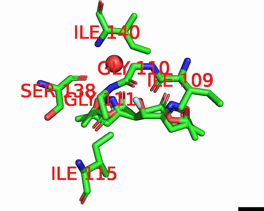
Mono view
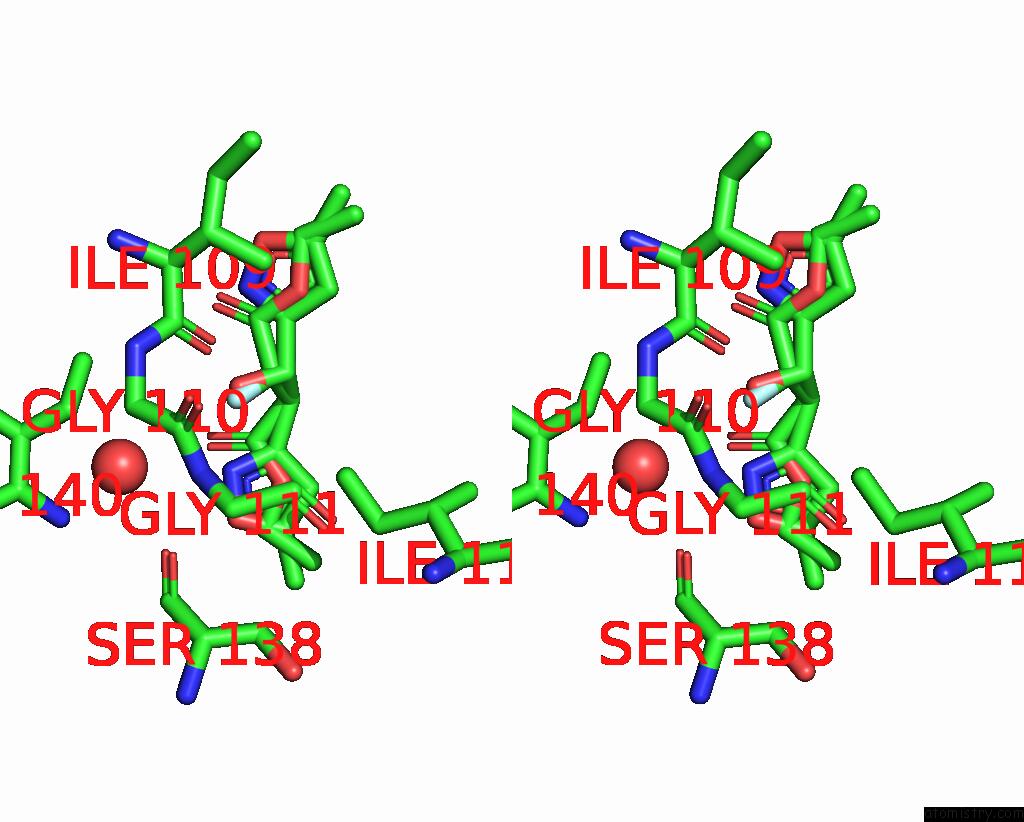
Stereo pair view
Mono view
Stereo pair view
A full contact list of Fluorine with other atoms in the F binding
site number 1 of Pandda Analysis Group Deposition -- Crystal Structure of Pseudomonas Aeruginosa Fabf-C164Q Mutant Protein in Complex with JKH100B within 5.0Å range:
|
Fluorine binding site 2 out of 2 in 5so3
Go back to
Fluorine binding site 2 out
of 2 in the Pandda Analysis Group Deposition -- Crystal Structure of Pseudomonas Aeruginosa Fabf-C164Q Mutant Protein in Complex with JKH100B
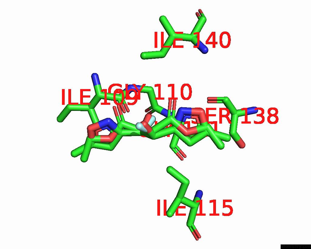
Mono view
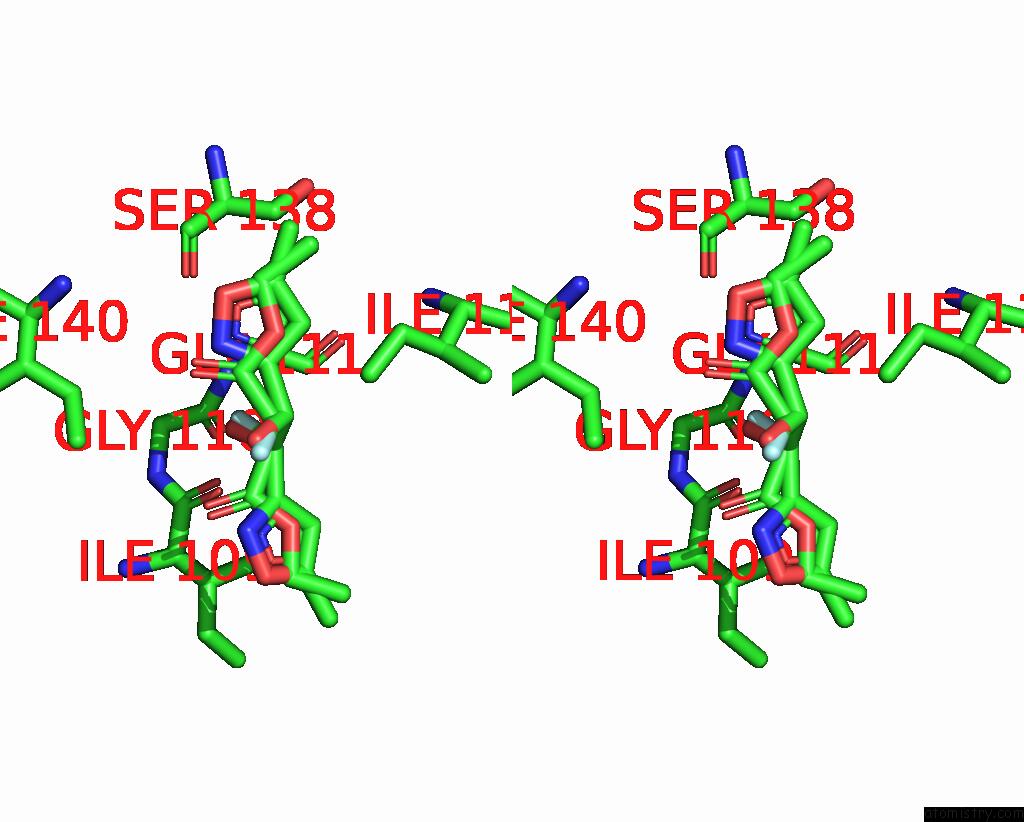
Stereo pair view
Mono view
Stereo pair view
A full contact list of Fluorine with other atoms in the F binding
site number 2 of Pandda Analysis Group Deposition -- Crystal Structure of Pseudomonas Aeruginosa Fabf-C164Q Mutant Protein in Complex with JKH100B within 5.0Å range:
|
Reference:
R.Brenk,
C.Georgiou.
Pandda Analysis Group Deposition To Be Published.
Page generated: Tue Jul 15 07:31:55 2025
Last articles
Mn in 5OXJMn in 5R7X
Mn in 5OX6
Mn in 5OX5
Mn in 5OR6
Mn in 5OVO
Mn in 5OR2
Mn in 5ONW
Mn in 5ONG
Mn in 5OLK