Fluorine »
PDB 5xdk-5y6d »
5xhc »
Fluorine in PDB 5xhc: Crystal Structure of T2R-Ttl-PO10 Complex
Protein crystallography data
The structure of Crystal Structure of T2R-Ttl-PO10 Complex, PDB code: 5xhc
was solved by
Y.Chu,
Y.Wang,
J.Yang,
W.Li,
with X-Ray Crystallography technique. A brief refinement statistics is given in the table below:
| Resolution Low / High (Å) | 119.38 / 2.75 |
| Space group | P 21 21 21 |
| Cell size a, b, c (Å), α, β, γ (°) | 105.257, 157.885, 182.427, 90.00, 90.00, 90.00 |
| R / Rfree (%) | 22.7 / 27.7 |
Other elements in 5xhc:
The structure of Crystal Structure of T2R-Ttl-PO10 Complex also contains other interesting chemical elements:
| Magnesium | (Mg) | 4 atoms |
| Calcium | (Ca) | 3 atoms |
Fluorine Binding Sites:
The binding sites of Fluorine atom in the Crystal Structure of T2R-Ttl-PO10 Complex
(pdb code 5xhc). This binding sites where shown within
5.0 Angstroms radius around Fluorine atom.
In total only one binding site of Fluorine was determined in the Crystal Structure of T2R-Ttl-PO10 Complex, PDB code: 5xhc:
In total only one binding site of Fluorine was determined in the Crystal Structure of T2R-Ttl-PO10 Complex, PDB code: 5xhc:
Fluorine binding site 1 out of 1 in 5xhc
Go back to
Fluorine binding site 1 out
of 1 in the Crystal Structure of T2R-Ttl-PO10 Complex
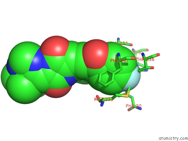
Mono view
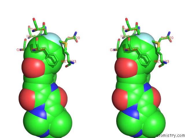
Stereo pair view
Mono view
Stereo pair view
A full contact list of Fluorine with other atoms in the F binding
site number 1 of Crystal Structure of T2R-Ttl-PO10 Complex within 5.0Å range:
|
Reference:
Y.Chu,
Y.Wang,
J.Yang,
W.Li.
Synthesis, Biological Evaluation and X-Ray Structure of Anti-Microtubule Agents To Be Published.
Page generated: Tue Jul 15 09:10:13 2025
Last articles
Na in 3M1HNa in 3M9Y
Na in 3M8A
Na in 3M92
Na in 3M1A
Na in 3M48
Na in 3M5P
Na in 3M3U
Na in 3M3M
Na in 3M1E