Fluorine »
PDB 6ffb-6g0l »
6fw9 »
Fluorine in PDB 6fw9: Crystal Structure of L-Tryptophan Oxidase Vioa From Chromobacterium Violaceum in Complex with 6-Fluoro-L-Tryptophan
Enzymatic activity of Crystal Structure of L-Tryptophan Oxidase Vioa From Chromobacterium Violaceum in Complex with 6-Fluoro-L-Tryptophan
All present enzymatic activity of Crystal Structure of L-Tryptophan Oxidase Vioa From Chromobacterium Violaceum in Complex with 6-Fluoro-L-Tryptophan:
1.4.3.23;
1.4.3.23;
Protein crystallography data
The structure of Crystal Structure of L-Tryptophan Oxidase Vioa From Chromobacterium Violaceum in Complex with 6-Fluoro-L-Tryptophan, PDB code: 6fw9
was solved by
H.E.Lai,
M.Morgan,
S.Moore,
P.Freemont,
with X-Ray Crystallography technique. A brief refinement statistics is given in the table below:
| Resolution Low / High (Å) | 63.86 / 2.74 |
| Space group | C 2 2 21 |
| Cell size a, b, c (Å), α, β, γ (°) | 151.478, 174.322, 93.821, 90.00, 90.00, 90.00 |
| R / Rfree (%) | 19.7 / 26.1 |
Other elements in 6fw9:
The structure of Crystal Structure of L-Tryptophan Oxidase Vioa From Chromobacterium Violaceum in Complex with 6-Fluoro-L-Tryptophan also contains other interesting chemical elements:
| Magnesium | (Mg) | 2 atoms |
Fluorine Binding Sites:
The binding sites of Fluorine atom in the Crystal Structure of L-Tryptophan Oxidase Vioa From Chromobacterium Violaceum in Complex with 6-Fluoro-L-Tryptophan
(pdb code 6fw9). This binding sites where shown within
5.0 Angstroms radius around Fluorine atom.
In total 3 binding sites of Fluorine where determined in the Crystal Structure of L-Tryptophan Oxidase Vioa From Chromobacterium Violaceum in Complex with 6-Fluoro-L-Tryptophan, PDB code: 6fw9:
Jump to Fluorine binding site number: 1; 2; 3;
In total 3 binding sites of Fluorine where determined in the Crystal Structure of L-Tryptophan Oxidase Vioa From Chromobacterium Violaceum in Complex with 6-Fluoro-L-Tryptophan, PDB code: 6fw9:
Jump to Fluorine binding site number: 1; 2; 3;
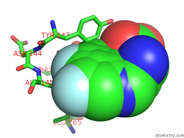
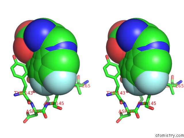
Fluorine binding site 1 out of 3 in 6fw9
Go back to
Fluorine binding site 1 out
of 3 in the Crystal Structure of L-Tryptophan Oxidase Vioa From Chromobacterium Violaceum in Complex with 6-Fluoro-L-Tryptophan
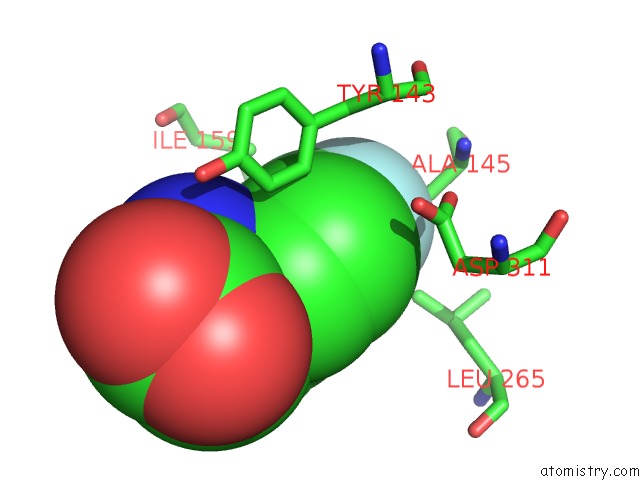
Mono view
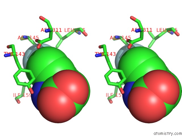
Stereo pair view
Mono view
Stereo pair view
A full contact list of Fluorine with other atoms in the F binding
site number 1 of Crystal Structure of L-Tryptophan Oxidase Vioa From Chromobacterium Violaceum in Complex with 6-Fluoro-L-Tryptophan within 5.0Å range:
|
Fluorine binding site 2 out of 3 in 6fw9
Go back to
Fluorine binding site 2 out
of 3 in the Crystal Structure of L-Tryptophan Oxidase Vioa From Chromobacterium Violaceum in Complex with 6-Fluoro-L-Tryptophan
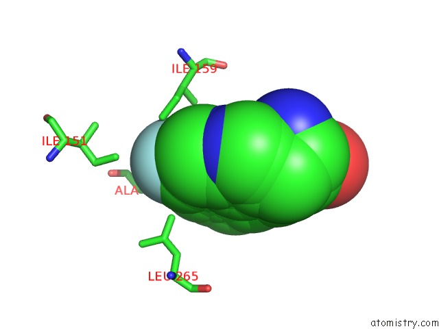
Mono view
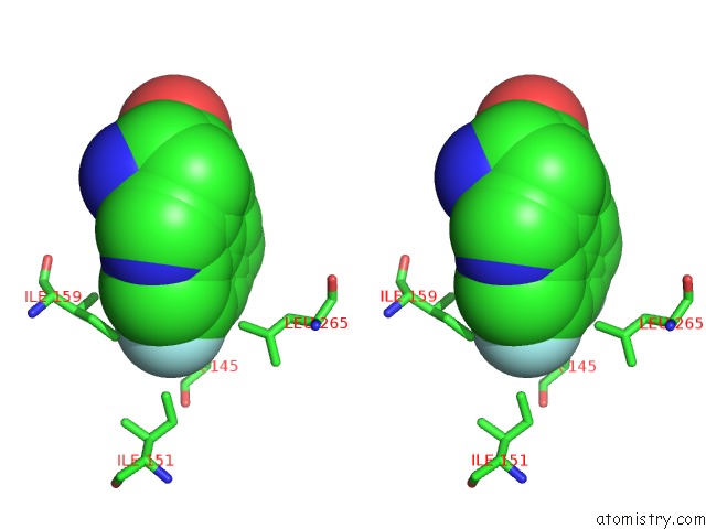
Stereo pair view
Mono view
Stereo pair view
A full contact list of Fluorine with other atoms in the F binding
site number 2 of Crystal Structure of L-Tryptophan Oxidase Vioa From Chromobacterium Violaceum in Complex with 6-Fluoro-L-Tryptophan within 5.0Å range:
|
Fluorine binding site 3 out of 3 in 6fw9
Go back to
Fluorine binding site 3 out
of 3 in the Crystal Structure of L-Tryptophan Oxidase Vioa From Chromobacterium Violaceum in Complex with 6-Fluoro-L-Tryptophan
Mono view
Stereo pair view
Mono view
Stereo pair view
A full contact list of Fluorine with other atoms in the F binding
site number 3 of Crystal Structure of L-Tryptophan Oxidase Vioa From Chromobacterium Violaceum in Complex with 6-Fluoro-L-Tryptophan within 5.0Å range:
|
Reference:
H.E.Lai,
A.M.C.Obled,
S.M.Chee,
R.M.Morgan,
S.V.Sharma,
S.J.Moore,
K.M.Polizzi,
R.J.M.Goss,
P.S.Freemont.
A Genochemetic Strategy For Derivatization of the Violacein Natural Product Scaffold Biorxiv 2019.
DOI: 10.1101/202523
Page generated: Tue Jul 15 11:41:37 2025
DOI: 10.1101/202523
Last articles
Mg in 5S61Mg in 5S60
Mg in 5S5Z
Mg in 5S5Y
Mg in 5S5X
Mg in 5S5W
Mg in 5S5V
Mg in 5S5U
Mg in 5S5T
Mg in 5S5S