Fluorine »
PDB 6hmr-6i78 »
6hob »
Fluorine in PDB 6hob: Transcriptional Repressor Ethr From Mycobacterium Tuberculosis in Complex with BDM44831
Protein crystallography data
The structure of Transcriptional Repressor Ethr From Mycobacterium Tuberculosis in Complex with BDM44831, PDB code: 6hob
was solved by
R.Wintjens,
A.Wohlkonig,
with X-Ray Crystallography technique. A brief refinement statistics is given in the table below:
| Resolution Low / High (Å) | 38.57 / 1.80 |
| Space group | P 41 21 2 |
| Cell size a, b, c (Å), α, β, γ (°) | 121.978, 121.978, 33.629, 90.00, 90.00, 90.00 |
| R / Rfree (%) | 18.3 / 21.3 |
Fluorine Binding Sites:
The binding sites of Fluorine atom in the Transcriptional Repressor Ethr From Mycobacterium Tuberculosis in Complex with BDM44831
(pdb code 6hob). This binding sites where shown within
5.0 Angstroms radius around Fluorine atom.
In total 3 binding sites of Fluorine where determined in the Transcriptional Repressor Ethr From Mycobacterium Tuberculosis in Complex with BDM44831, PDB code: 6hob:
Jump to Fluorine binding site number: 1; 2; 3;
In total 3 binding sites of Fluorine where determined in the Transcriptional Repressor Ethr From Mycobacterium Tuberculosis in Complex with BDM44831, PDB code: 6hob:
Jump to Fluorine binding site number: 1; 2; 3;
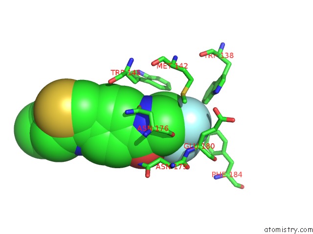
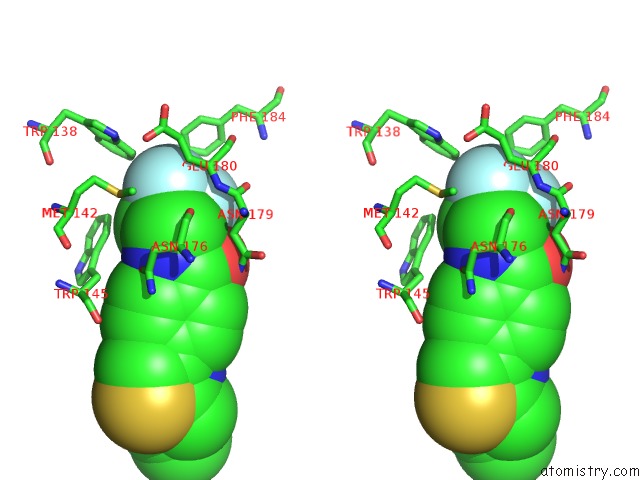
Fluorine binding site 1 out of 3 in 6hob
Go back to
Fluorine binding site 1 out
of 3 in the Transcriptional Repressor Ethr From Mycobacterium Tuberculosis in Complex with BDM44831
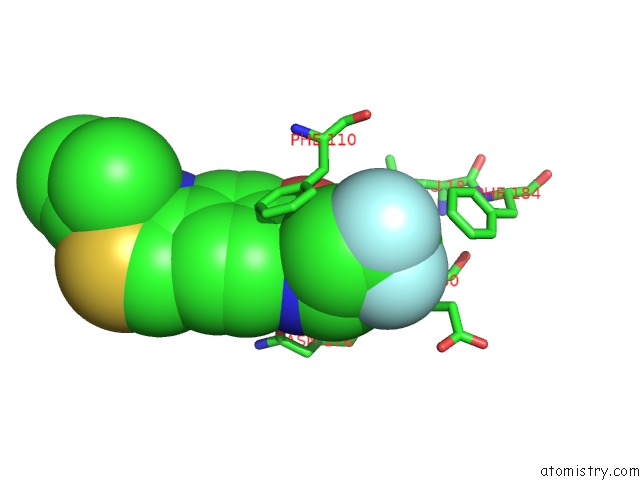
Mono view
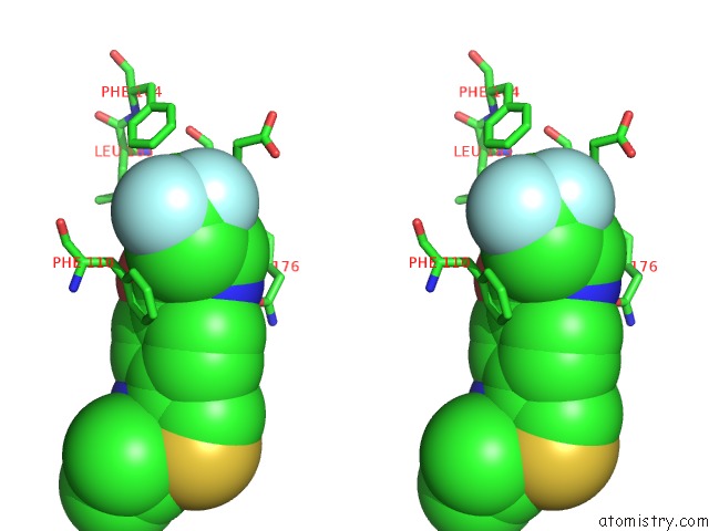
Stereo pair view
Mono view
Stereo pair view
A full contact list of Fluorine with other atoms in the F binding
site number 1 of Transcriptional Repressor Ethr From Mycobacterium Tuberculosis in Complex with BDM44831 within 5.0Å range:
|
Fluorine binding site 2 out of 3 in 6hob
Go back to
Fluorine binding site 2 out
of 3 in the Transcriptional Repressor Ethr From Mycobacterium Tuberculosis in Complex with BDM44831
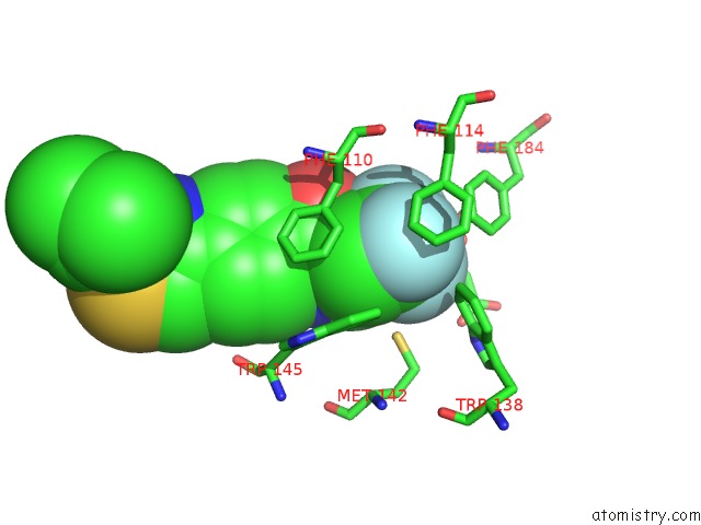
Mono view
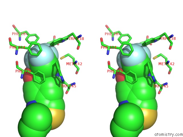
Stereo pair view
Mono view
Stereo pair view
A full contact list of Fluorine with other atoms in the F binding
site number 2 of Transcriptional Repressor Ethr From Mycobacterium Tuberculosis in Complex with BDM44831 within 5.0Å range:
|
Fluorine binding site 3 out of 3 in 6hob
Go back to
Fluorine binding site 3 out
of 3 in the Transcriptional Repressor Ethr From Mycobacterium Tuberculosis in Complex with BDM44831
Mono view
Stereo pair view
Mono view
Stereo pair view
A full contact list of Fluorine with other atoms in the F binding
site number 3 of Transcriptional Repressor Ethr From Mycobacterium Tuberculosis in Complex with BDM44831 within 5.0Å range:
|
Reference:
A.Tanina,
A.Wohlkonig,
S.H.Soror,
M.Flipo,
B.Villemagne,
H.Prevet,
B.Deprez,
M.Moune,
H.Peree,
F.Meyer,
A.R.Baulard,
N.Willand,
R.Wintjens.
A Comprehensive Analysis of the Protein-Ligand Interactions in Crystal Structures of Mycobacterium Tuberculosis Ethr. Biochim Biophys Acta V.1867 248 2018PROTEINS Proteom.
ISSN: ISSN 1878-1454
PubMed: 30553830
DOI: 10.1016/J.BBAPAP.2018.12.003
Page generated: Tue Jul 15 12:23:44 2025
ISSN: ISSN 1878-1454
PubMed: 30553830
DOI: 10.1016/J.BBAPAP.2018.12.003
Last articles
Mg in 4ZOBMg in 4ZOA
Mg in 4ZO9
Mg in 4ZO8
Mg in 4ZO7
Mg in 4ZO6
Mg in 4ZO5
Mg in 4ZO0
Mg in 4ZNP
Mg in 4ZNL