Fluorine »
PDB 6i8z-6jsf »
6il0 »
Fluorine in PDB 6il0: K3U Complex Structure of Peptide Deformylase From Xanthomonas Oryzae Pv. Oryzae
Enzymatic activity of K3U Complex Structure of Peptide Deformylase From Xanthomonas Oryzae Pv. Oryzae
All present enzymatic activity of K3U Complex Structure of Peptide Deformylase From Xanthomonas Oryzae Pv. Oryzae:
3.5.1.88;
3.5.1.88;
Protein crystallography data
The structure of K3U Complex Structure of Peptide Deformylase From Xanthomonas Oryzae Pv. Oryzae, PDB code: 6il0
was solved by
I.H.Lee,
L.W.Kang,
with X-Ray Crystallography technique. A brief refinement statistics is given in the table below:
| Resolution Low / High (Å) | 49.91 / 1.93 |
| Space group | P 61 2 2 |
| Cell size a, b, c (Å), α, β, γ (°) | 58.682, 58.682, 265.531, 90.00, 90.00, 120.00 |
| R / Rfree (%) | 19.5 / 23.1 |
Other elements in 6il0:
The structure of K3U Complex Structure of Peptide Deformylase From Xanthomonas Oryzae Pv. Oryzae also contains other interesting chemical elements:
| Nickel | (Ni) | 3 atoms |
Fluorine Binding Sites:
The binding sites of Fluorine atom in the K3U Complex Structure of Peptide Deformylase From Xanthomonas Oryzae Pv. Oryzae
(pdb code 6il0). This binding sites where shown within
5.0 Angstroms radius around Fluorine atom.
In total 3 binding sites of Fluorine where determined in the K3U Complex Structure of Peptide Deformylase From Xanthomonas Oryzae Pv. Oryzae, PDB code: 6il0:
Jump to Fluorine binding site number: 1; 2; 3;
In total 3 binding sites of Fluorine where determined in the K3U Complex Structure of Peptide Deformylase From Xanthomonas Oryzae Pv. Oryzae, PDB code: 6il0:
Jump to Fluorine binding site number: 1; 2; 3;
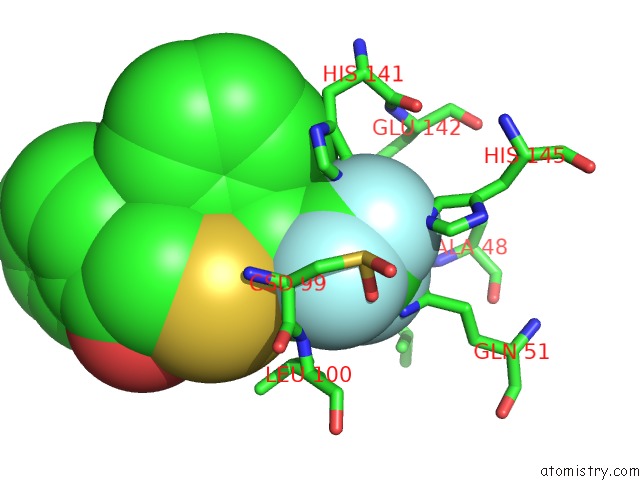
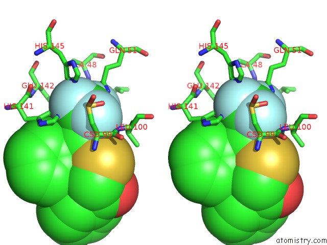
Fluorine binding site 1 out of 3 in 6il0
Go back to
Fluorine binding site 1 out
of 3 in the K3U Complex Structure of Peptide Deformylase From Xanthomonas Oryzae Pv. Oryzae
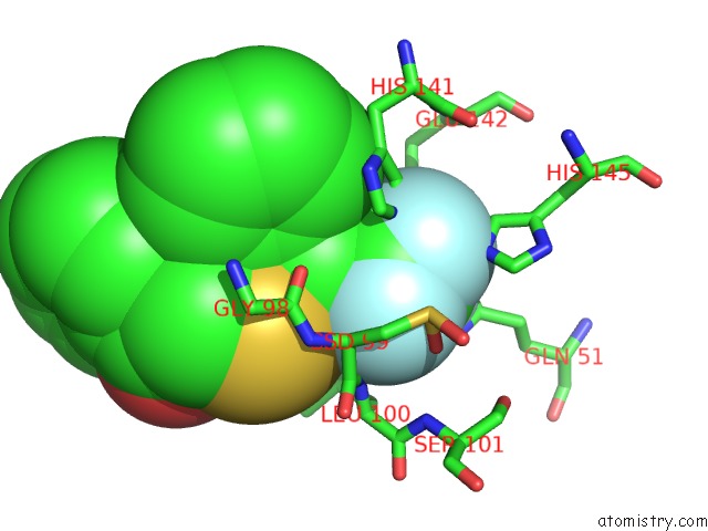
Mono view
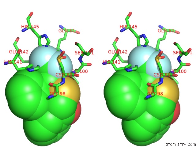
Stereo pair view
Mono view
Stereo pair view
A full contact list of Fluorine with other atoms in the F binding
site number 1 of K3U Complex Structure of Peptide Deformylase From Xanthomonas Oryzae Pv. Oryzae within 5.0Å range:
|
Fluorine binding site 2 out of 3 in 6il0
Go back to
Fluorine binding site 2 out
of 3 in the K3U Complex Structure of Peptide Deformylase From Xanthomonas Oryzae Pv. Oryzae
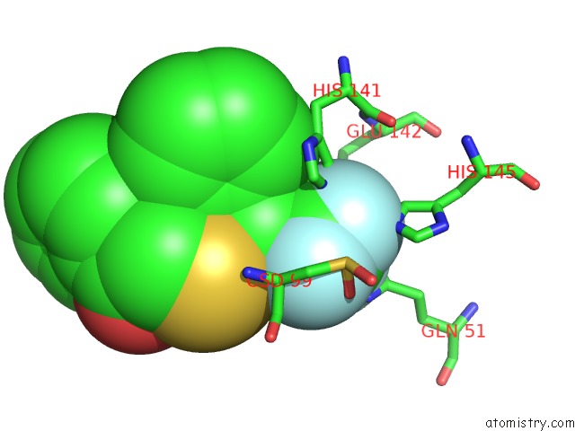
Mono view
Stereo pair view
Mono view
Stereo pair view
A full contact list of Fluorine with other atoms in the F binding
site number 2 of K3U Complex Structure of Peptide Deformylase From Xanthomonas Oryzae Pv. Oryzae within 5.0Å range:
|
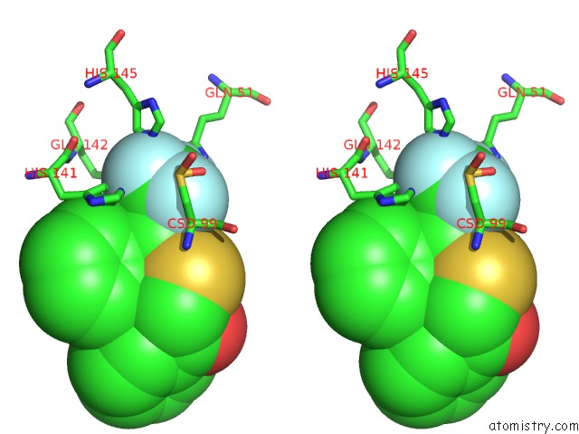
Fluorine binding site 3 out of 3 in 6il0
Go back to
Fluorine binding site 3 out
of 3 in the K3U Complex Structure of Peptide Deformylase From Xanthomonas Oryzae Pv. Oryzae
Mono view
Stereo pair view
Mono view
Stereo pair view
A full contact list of Fluorine with other atoms in the F binding
site number 3 of K3U Complex Structure of Peptide Deformylase From Xanthomonas Oryzae Pv. Oryzae within 5.0Å range:
|
Reference:
I.H.Lee,
L.W.Kang.
Fbis Complex Structure of Peptide Deformylase From Xanthomonas Oryzae Pv. Oryzae To Be Published.
Page generated: Tue Jul 15 12:30:44 2025
Last articles
Mg in 5EX5Mg in 5EX2
Mg in 5EX1
Mg in 5EW7
Mg in 5EW4
Mg in 5EVZ
Mg in 5ETS
Mg in 5ETT
Mg in 5EUR
Mg in 5EUL