Atomistry » Fluorine » PDB 6i8z-6jsf » 6jf7
Atomistry »
  Fluorine »
    PDB 6i8z-6jsf »
      6jf7 »

Fluorine in PDB 6jf7: K3U Bound Crystal Structure of Class I Type B Peptide Deformylase From Acinetobacter Baumannii

Enzymatic activity of K3U Bound Crystal Structure of Class I Type B Peptide Deformylase From Acinetobacter Baumannii

All present enzymatic activity of K3U Bound Crystal Structure of Class I Type B Peptide Deformylase From Acinetobacter Baumannii:
3.5.1.88;

Protein crystallography data

The structure of K3U Bound Crystal Structure of Class I Type B Peptide Deformylase From Acinetobacter Baumannii, PDB code: 6jf7 was solved by K.H.Jung, T.H.Ho, I.H.Lee, L.W.Kang, with X-Ray Crystallography technique. A brief refinement statistics is given in the table below:

Resolution Low / High (Å) 49.99 / 2.00
Space group P 21 21 21
Cell size a, b, c (Å), α, β, γ (°) 39.513, 71.259, 110.974, 90.00, 90.00, 90.00
R / Rfree (%) 19.2 / 24.8

Other elements in 6jf7:

The structure of K3U Bound Crystal Structure of Class I Type B Peptide Deformylase From Acinetobacter Baumannii also contains other interesting chemical elements:

Zinc (Zn) 2 atoms

Fluorine Binding Sites:

The binding sites of Fluorine atom in the K3U Bound Crystal Structure of Class I Type B Peptide Deformylase From Acinetobacter Baumannii (pdb code 6jf7). This binding sites where shown within 5.0 Angstroms radius around Fluorine atom.
In total 6 binding sites of Fluorine where determined in the K3U Bound Crystal Structure of Class I Type B Peptide Deformylase From Acinetobacter Baumannii, PDB code: 6jf7:
Jump to Fluorine binding site number: 1; 2; 3; 4; 5; 6;

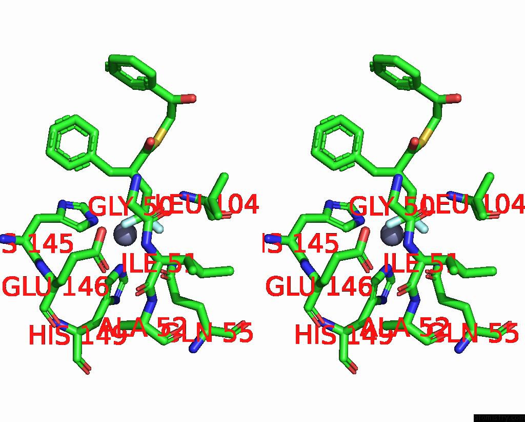
Fluorine binding site 1 out of 6 in 6jf7

Go back to Fluorine Binding Sites List in 6jf7
Fluorine binding site 1 out of 6 in the K3U Bound Crystal Structure of Class I Type B Peptide Deformylase From Acinetobacter Baumannii


Mono view


Stereo pair view

A full contact list of Fluorine with other atoms in the F binding site number 1 of K3U Bound Crystal Structure of Class I Type B Peptide Deformylase From Acinetobacter Baumannii within 5.0Å range:
probe atom residue distance (Å) B Occ
A:F202

b:29.6
occ:1.00
F1 A:K3U202 0.0 29.6 1.0
C12 A:K3U202 1.3 33.8 1.0
ZN A:ZN201 1.9 20.1 1.0
F A:K3U202 2.1 33.2 1.0
F2 A:K3U202 2.1 64.3 1.0
C11 A:K3U202 2.4 35.6 1.0
NE2 A:GLN55 3.1 18.0 1.0
C3 A:K3U202 3.1 33.2 1.0
N A:LEU104 3.1 20.6 1.0
NE2 A:HIS145 3.2 16.0 1.0
SG A:CYS103 3.2 20.3 1.0
CA A:CYS103 3.2 20.3 1.0
NE2 A:HIS149 3.5 17.4 1.0
CB A:CYS103 3.6 21.1 1.0
C A:CYS103 3.7 21.0 1.0
OE1 A:GLN55 3.8 18.3 1.0
CD A:GLN55 3.8 17.5 1.0
CD2 A:HIS145 3.9 15.5 1.0
C4 A:K3U202 4.1 31.1 1.0
CE1 A:HIS145 4.1 19.2 1.0
CG A:LEU104 4.2 27.1 1.0
C2 A:K3U202 4.2 39.6 1.0
CA A:LEU104 4.2 22.1 1.0
CB A:LEU104 4.3 24.5 1.0
O A:GLY102 4.3 21.7 1.0
CD2 A:HIS149 4.4 16.0 1.0
OE2 A:GLU146 4.5 21.0 1.0
N A:CYS103 4.5 20.9 1.0
S A:K3U202 4.5 45.2 1.0
CE1 A:HIS149 4.5 17.1 1.0
N A:SER105 4.7 20.2 1.0
CD1 A:LEU104 4.7 28.5 1.0
C A:GLY102 4.8 20.7 1.0
O A:CYS103 4.9 18.3 1.0
O A:GLY50 4.9 17.8 1.0
O A:HOH311 5.0 20.9 1.0
OE1 A:GLU146 5.0 16.7 1.0

Fluorine binding site 2 out of 6 in 6jf7

Go back to Fluorine Binding Sites List in 6jf7
Fluorine binding site 2 out of 6 in the K3U Bound Crystal Structure of Class I Type B Peptide Deformylase From Acinetobacter Baumannii


Mono view


Stereo pair view

A full contact list of Fluorine with other atoms in the F binding site number 2 of K3U Bound Crystal Structure of Class I Type B Peptide Deformylase From Acinetobacter Baumannii within 5.0Å range:
probe atom residue distance (Å) B Occ
A:F202

b:33.2
occ:1.00
F A:K3U202 0.0 33.2 1.0
C12 A:K3U202 1.3 33.8 1.0
F2 A:K3U202 2.0 64.3 1.0
F1 A:K3U202 2.1 29.6 1.0
C11 A:K3U202 2.3 35.6 1.0
OE2 A:GLU146 2.4 21.0 1.0
ZN A:ZN201 2.8 20.1 1.0
CD2 A:HIS145 3.0 15.5 1.0
C3 A:K3U202 3.1 33.2 1.0
NE2 A:HIS145 3.1 16.0 1.0
CD A:GLU146 3.1 19.1 1.0
C4 A:K3U202 3.2 31.1 1.0
OE1 A:GLU146 3.2 16.7 1.0
OE1 A:GLN55 3.6 18.3 1.0
NE2 A:HIS149 3.7 17.4 1.0
CD2 A:HIS149 3.9 16.0 1.0
O A:GLY50 3.9 17.8 1.0
NE2 A:GLN55 4.1 18.0 1.0
C A:GLY50 4.2 16.4 1.0
CG A:HIS145 4.2 16.0 1.0
CE1 A:HIS145 4.2 19.2 1.0
CD A:GLN55 4.3 17.5 1.0
CA A:ILE51 4.4 15.9 1.0
C5 A:K3U202 4.4 29.7 1.0
N A:ILE51 4.4 15.5 1.0
C2 A:K3U202 4.5 39.6 1.0
CG A:GLU146 4.6 17.2 1.0
C10 A:K3U202 4.7 29.2 1.0
ND1 A:HIS145 4.8 17.1 1.0
CA A:GLY50 4.8 17.8 1.0
SG A:CYS103 4.9 20.3 1.0

Fluorine binding site 3 out of 6 in 6jf7

Go back to Fluorine Binding Sites List in 6jf7
Fluorine binding site 3 out of 6 in the K3U Bound Crystal Structure of Class I Type B Peptide Deformylase From Acinetobacter Baumannii


Mono view


Stereo pair view

A full contact list of Fluorine with other atoms in the F binding site number 3 of K3U Bound Crystal Structure of Class I Type B Peptide Deformylase From Acinetobacter Baumannii within 5.0Å range:
probe atom residue distance (Å) B Occ
A:F202

b:64.3
occ:1.00
F2 A:K3U202 0.0 64.3 1.0
C12 A:K3U202 1.3 33.8 1.0
F A:K3U202 2.0 33.2 1.0
F1 A:K3U202 2.1 29.6 1.0
C11 A:K3U202 2.3 35.6 1.0
OE1 A:GLN55 2.6 18.3 1.0
NE2 A:GLN55 3.1 18.0 1.0
O A:GLY50 3.2 17.8 1.0
CD A:GLN55 3.2 17.5 1.0
CD1 A:LEU104 3.5 28.5 1.0
ZN A:ZN201 3.6 20.1 1.0
CG A:LEU104 3.6 27.1 1.0
CA A:ILE51 3.7 15.9 1.0
C3 A:K3U202 3.7 33.2 1.0
OE2 A:GLU146 3.7 21.0 1.0
C A:GLY50 3.9 16.4 1.0
OE1 A:GLU146 4.1 16.7 1.0
N A:ILE51 4.1 15.5 1.0
CB A:LEU104 4.2 24.5 1.0
N A:LEU104 4.3 20.6 1.0
CB A:ILE51 4.3 16.3 1.0
CG1 A:ILE51 4.3 17.1 1.0
NE2 A:HIS149 4.3 17.4 1.0
CD A:GLU146 4.3 19.1 1.0
C4 A:K3U202 4.4 31.1 1.0
C2 A:K3U202 4.6 39.6 1.0
CD2 A:HIS149 4.6 16.0 1.0
C A:ILE51 4.7 14.9 1.0
N A:ALA52 4.7 14.2 1.0
NE2 A:HIS145 4.7 16.0 1.0
CG A:GLN55 4.7 16.6 1.0
CA A:LEU104 4.9 22.1 1.0
CD2 A:HIS145 4.9 15.5 1.0
CD2 A:LEU104 5.0 31.9 1.0

Fluorine binding site 4 out of 6 in 6jf7

Go back to Fluorine Binding Sites List in 6jf7
Fluorine binding site 4 out of 6 in the K3U Bound Crystal Structure of Class I Type B Peptide Deformylase From Acinetobacter Baumannii


Mono view


Stereo pair view

A full contact list of Fluorine with other atoms in the F binding site number 4 of K3U Bound Crystal Structure of Class I Type B Peptide Deformylase From Acinetobacter Baumannii within 5.0Å range:
probe atom residue distance (Å) B Occ
B:F202

b:62.8
occ:1.00
F1 B:K3U202 0.0 62.8 1.0
C12 B:K3U202 1.3 36.2 1.0
C11 B:K3U202 2.1 36.5 1.0
F B:K3U202 2.2 28.4 1.0
F2 B:K3U202 2.2 28.6 1.0
NE2 B:GLN55 3.0 17.5 1.0
OE1 B:GLN55 3.1 19.4 1.0
CD1 B:LEU104 3.2 34.7 1.0
O B:GLY50 3.2 17.4 1.0
CG B:LEU104 3.2 32.6 1.0
CD B:GLN55 3.5 17.6 1.0
C3 B:K3U202 3.6 38.2 1.0
ZN B:ZN201 3.6 20.7 1.0
CB B:LEU104 3.7 29.6 1.0
N B:LEU104 4.0 25.8 1.0
OE2 B:GLU146 4.1 19.8 1.0
C B:GLY50 4.1 15.7 1.0
CA B:ILE51 4.2 14.6 1.0
C2 B:K3U202 4.3 42.3 1.0
C4 B:K3U202 4.4 35.9 1.0
CA B:LEU104 4.5 27.6 1.0
OE1 B:GLU146 4.5 20.3 1.0
N B:ILE51 4.6 14.4 1.0
CD2 B:LEU104 4.6 37.0 1.0
CB B:ILE51 4.7 13.8 1.0
CG1 B:ILE51 4.7 14.7 1.0
NE2 B:HIS149 4.7 15.5 1.0
CD B:GLU146 4.7 19.9 1.0
O1 B:K3U202 4.9 37.8 1.0
NE2 B:HIS145 4.9 21.1 1.0
CG B:GLN55 4.9 16.3 1.0
S B:K3U202 5.0 61.1 1.0
SG B:CYS103 5.0 25.6 1.0
CD2 B:HIS149 5.0 16.5 1.0

Fluorine binding site 5 out of 6 in 6jf7

Go back to Fluorine Binding Sites List in 6jf7
Fluorine binding site 5 out of 6 in the K3U Bound Crystal Structure of Class I Type B Peptide Deformylase From Acinetobacter Baumannii


Mono view


Stereo pair view

A full contact list of Fluorine with other atoms in the F binding site number 5 of K3U Bound Crystal Structure of Class I Type B Peptide Deformylase From Acinetobacter Baumannii within 5.0Å range:
probe atom residue distance (Å) B Occ
B:F202

b:28.4
occ:1.00
F B:K3U202 0.0 28.4 1.0
C12 B:K3U202 1.3 36.2 1.0
F1 B:K3U202 2.2 62.8 1.0
F2 B:K3U202 2.2 28.6 1.0
C11 B:K3U202 2.2 36.5 1.0
OE2 B:GLU146 2.4 19.8 1.0
ZN B:ZN201 2.8 20.7 1.0
CD B:GLU146 3.1 19.9 1.0
OE1 B:GLU146 3.1 20.3 1.0
C3 B:K3U202 3.2 38.2 1.0
CD2 B:HIS145 3.2 19.5 1.0
NE2 B:HIS145 3.3 21.1 1.0
C4 B:K3U202 3.4 35.9 1.0
OE1 B:GLN55 3.5 19.4 1.0
O B:GLY50 3.7 17.4 1.0
CD2 B:HIS149 3.8 16.5 1.0
NE2 B:HIS149 3.8 15.5 1.0
NE2 B:GLN55 3.9 17.5 1.0
CD B:GLN55 4.1 17.6 1.0
C B:GLY50 4.1 15.7 1.0
CA B:ILE51 4.4 14.6 1.0
CG B:HIS145 4.4 18.6 1.0
N B:ILE51 4.4 14.4 1.0
CE1 B:HIS145 4.5 21.2 1.0
CG B:GLU146 4.5 17.4 1.0
C2 B:K3U202 4.5 42.3 1.0
C5 B:K3U202 4.6 33.5 1.0
CA B:GLY50 4.9 15.8 1.0
SG B:CYS103 4.9 25.6 1.0
C10 B:K3U202 5.0 33.8 1.0

Fluorine binding site 6 out of 6 in 6jf7

Go back to Fluorine Binding Sites List in 6jf7
Fluorine binding site 6 out of 6 in the K3U Bound Crystal Structure of Class I Type B Peptide Deformylase From Acinetobacter Baumannii


Mono view


Stereo pair view

A full contact list of Fluorine with other atoms in the F binding site number 6 of K3U Bound Crystal Structure of Class I Type B Peptide Deformylase From Acinetobacter Baumannii within 5.0Å range:
probe atom residue distance (Å) B Occ
B:F202

b:28.6
occ:1.00
F2 B:K3U202 0.0 28.6 1.0
C12 B:K3U202 1.3 36.2 1.0
ZN B:ZN201 1.9 20.7 1.0
F1 B:K3U202 2.2 62.8 1.0
F B:K3U202 2.2 28.4 1.0
C11 B:K3U202 2.4 36.5 1.0
C3 B:K3U202 3.1 38.2 1.0
SG B:CYS103 3.1 25.6 1.0
NE2 B:HIS145 3.1 21.1 1.0
N B:LEU104 3.2 25.8 1.0
NE2 B:GLN55 3.2 17.5 1.0
CA B:CYS103 3.2 27.2 1.0
CB B:CYS103 3.6 27.5 1.0
NE2 B:HIS149 3.6 15.5 1.0
C B:CYS103 3.7 28.7 1.0
CD2 B:HIS145 3.9 19.5 1.0
CD B:GLN55 4.0 17.6 1.0
CE1 B:HIS145 4.1 21.2 1.0
C4 B:K3U202 4.1 35.9 1.0
OE1 B:GLN55 4.1 19.4 1.0
C2 B:K3U202 4.1 42.3 1.0
CB B:LEU104 4.2 29.6 1.0
CA B:LEU104 4.2 27.6 1.0
CG B:LEU104 4.3 32.6 1.0
O B:GLY102 4.3 25.3 1.0
CD2 B:HIS149 4.4 16.5 1.0
N B:CYS103 4.4 28.6 1.0
S B:K3U202 4.4 61.1 1.0
N B:SER105 4.5 24.4 1.0
OE2 B:GLU146 4.6 19.8 1.0
CE1 B:HIS149 4.7 16.0 1.0
C B:GLY102 4.8 28.1 1.0
CD1 B:LEU104 4.8 34.7 1.0
C B:LEU104 4.9 27.1 1.0
O B:CYS103 4.9 27.9 1.0
O B:HOH315 4.9 24.0 1.0

Reference:

K.H.Jung, T.H.Ho, I.H.Lee, L.W.Kang. K3U Bound Crystal Structure of Class I Type B Peptide Deformylase From Acinetobacter Baumannii To Be Published.
Page generated: Tue Jul 15 12:38:29 2025

Last articles

Fe in 2YXO
Fe in 2YRS
Fe in 2YXC
Fe in 2YNM
Fe in 2YVJ
Fe in 2YP1
Fe in 2YU2
Fe in 2YU1
Fe in 2YQB
Fe in 2YOO
© Copyright 2008-2020 by atomistry.com
Home   |    Site Map   |    Copyright   |    Contact us   |    Privacy