Fluorine »
PDB 6i8z-6jsf »
6jfe »
Fluorine in PDB 6jfe: K2U Bound Crystal Structure of Class I Type B Peptide Deformylase From Pseudomonas Aeruginosa
Enzymatic activity of K2U Bound Crystal Structure of Class I Type B Peptide Deformylase From Pseudomonas Aeruginosa
All present enzymatic activity of K2U Bound Crystal Structure of Class I Type B Peptide Deformylase From Pseudomonas Aeruginosa:
3.5.1.88;
3.5.1.88;
Protein crystallography data
The structure of K2U Bound Crystal Structure of Class I Type B Peptide Deformylase From Pseudomonas Aeruginosa, PDB code: 6jfe
was solved by
I.H.Lee,
T.H.Ho,
L.W.Kang,
with X-Ray Crystallography technique. A brief refinement statistics is given in the table below:
| Resolution Low / High (Å) | 46.81 / 2.10 |
| Space group | C 2 2 21 |
| Cell size a, b, c (Å), α, β, γ (°) | 44.682, 121.818, 146.314, 90.00, 90.00, 90.00 |
| R / Rfree (%) | 20.7 / 25.2 |
Other elements in 6jfe:
The structure of K2U Bound Crystal Structure of Class I Type B Peptide Deformylase From Pseudomonas Aeruginosa also contains other interesting chemical elements:
| Nickel | (Ni) | 4 atoms |
Fluorine Binding Sites:
The binding sites of Fluorine atom in the K2U Bound Crystal Structure of Class I Type B Peptide Deformylase From Pseudomonas Aeruginosa
(pdb code 6jfe). This binding sites where shown within
5.0 Angstroms radius around Fluorine atom.
In total 3 binding sites of Fluorine where determined in the K2U Bound Crystal Structure of Class I Type B Peptide Deformylase From Pseudomonas Aeruginosa, PDB code: 6jfe:
Jump to Fluorine binding site number: 1; 2; 3;
In total 3 binding sites of Fluorine where determined in the K2U Bound Crystal Structure of Class I Type B Peptide Deformylase From Pseudomonas Aeruginosa, PDB code: 6jfe:
Jump to Fluorine binding site number: 1; 2; 3;
Fluorine binding site 1 out of 3 in 6jfe
Go back to
Fluorine binding site 1 out
of 3 in the K2U Bound Crystal Structure of Class I Type B Peptide Deformylase From Pseudomonas Aeruginosa
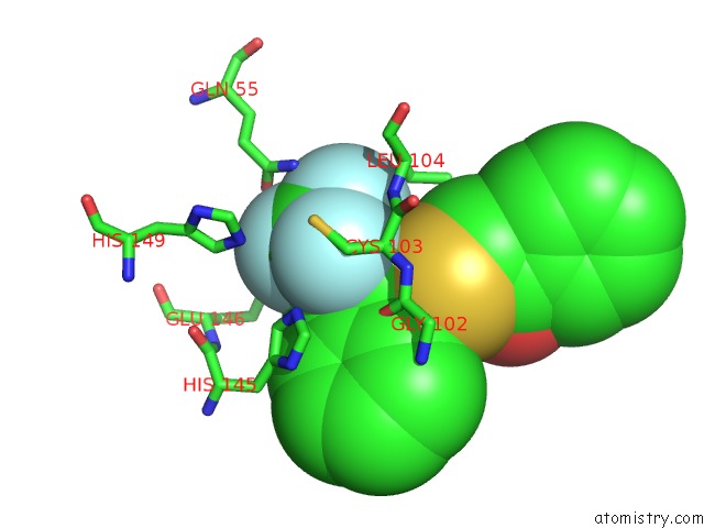
Mono view

Stereo pair view
Mono view
Stereo pair view
A full contact list of Fluorine with other atoms in the F binding
site number 1 of K2U Bound Crystal Structure of Class I Type B Peptide Deformylase From Pseudomonas Aeruginosa within 5.0Å range:
|
Fluorine binding site 2 out of 3 in 6jfe
Go back to
Fluorine binding site 2 out
of 3 in the K2U Bound Crystal Structure of Class I Type B Peptide Deformylase From Pseudomonas Aeruginosa
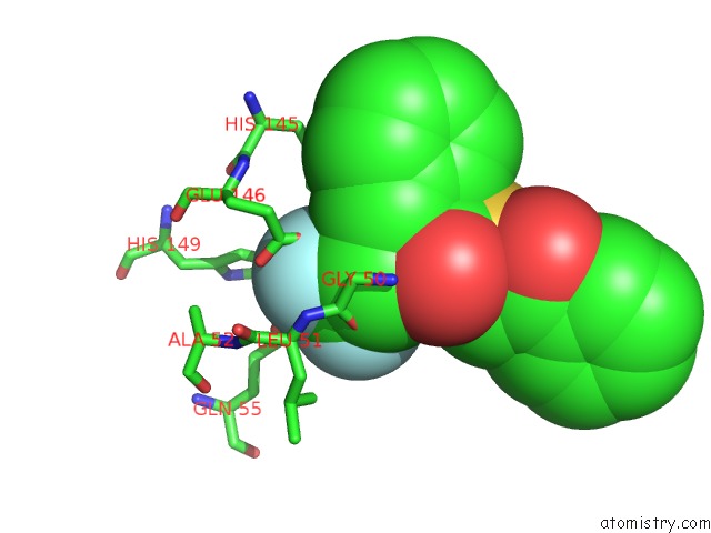
Mono view

Stereo pair view
Mono view
Stereo pair view
A full contact list of Fluorine with other atoms in the F binding
site number 2 of K2U Bound Crystal Structure of Class I Type B Peptide Deformylase From Pseudomonas Aeruginosa within 5.0Å range:
|
Fluorine binding site 3 out of 3 in 6jfe
Go back to
Fluorine binding site 3 out
of 3 in the K2U Bound Crystal Structure of Class I Type B Peptide Deformylase From Pseudomonas Aeruginosa
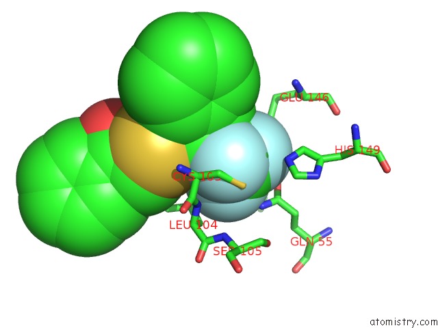
Mono view
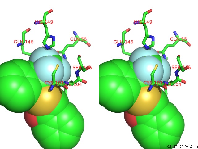
Stereo pair view
Mono view
Stereo pair view
A full contact list of Fluorine with other atoms in the F binding
site number 3 of K2U Bound Crystal Structure of Class I Type B Peptide Deformylase From Pseudomonas Aeruginosa within 5.0Å range:
|
Reference:
I.H.Lee,
T.H.Ho,
L.W.Kang.
K2U Bound Crystal Structure of Class I Type B Peptide Deformylase From Pseudomonas Aeruginosa To Be Published.
Page generated: Tue Jul 15 12:39:02 2025
Last articles
Mg in 5FTNMg in 5FUX
Mg in 5FUK
Mg in 5FTM
Mg in 5FUJ
Mg in 5FUI
Mg in 5FTE
Mg in 5FTB
Mg in 5FSC
Mg in 5FSB