Fluorine »
PDB 6i8z-6jsf »
6jok »
Fluorine in PDB 6jok: Crystal Structure of Pdgfra in Complex with Sunitinib By Soaking
Enzymatic activity of Crystal Structure of Pdgfra in Complex with Sunitinib By Soaking
All present enzymatic activity of Crystal Structure of Pdgfra in Complex with Sunitinib By Soaking:
2.7.10.1;
2.7.10.1;
Protein crystallography data
The structure of Crystal Structure of Pdgfra in Complex with Sunitinib By Soaking, PDB code: 6jok
was solved by
L.Liang,
X.E.Yan,
C.H.Yun,
with X-Ray Crystallography technique. A brief refinement statistics is given in the table below:
| Resolution Low / High (Å) | 47.17 / 3.80 |
| Space group | P 31 2 1 |
| Cell size a, b, c (Å), α, β, γ (°) | 102.611, 102.611, 111.301, 90.00, 90.00, 120.00 |
| R / Rfree (%) | 25.7 / 28.2 |
Other elements in 6jok:
The structure of Crystal Structure of Pdgfra in Complex with Sunitinib By Soaking also contains other interesting chemical elements:
| Chlorine | (Cl) | 2 atoms |
Fluorine Binding Sites:
The binding sites of Fluorine atom in the Crystal Structure of Pdgfra in Complex with Sunitinib By Soaking
(pdb code 6jok). This binding sites where shown within
5.0 Angstroms radius around Fluorine atom.
In total only one binding site of Fluorine was determined in the Crystal Structure of Pdgfra in Complex with Sunitinib By Soaking, PDB code: 6jok:
In total only one binding site of Fluorine was determined in the Crystal Structure of Pdgfra in Complex with Sunitinib By Soaking, PDB code: 6jok:
Fluorine binding site 1 out of 1 in 6jok
Go back to
Fluorine binding site 1 out
of 1 in the Crystal Structure of Pdgfra in Complex with Sunitinib By Soaking
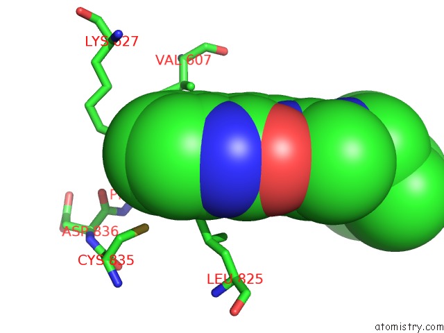
Mono view
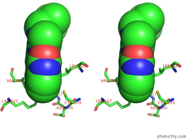
Stereo pair view
Mono view
Stereo pair view
A full contact list of Fluorine with other atoms in the F binding
site number 1 of Crystal Structure of Pdgfra in Complex with Sunitinib By Soaking within 5.0Å range:
|
Reference:
L.Liang,
X.E.Yan.
Crystal Structure of Pdgfra in Complex with Sunitinib By Soaking To Be Published.
Page generated: Tue Jul 15 12:41:23 2025
Last articles
Mg in 5ZKJMg in 5ZKI
Mg in 5ZK6
Mg in 5ZE9
Mg in 5ZFX
Mg in 5ZCT
Mg in 5ZE6
Mg in 5ZE4
Mg in 5ZDN
Mg in 5ZE0