Fluorine »
PDB 6jsg-6kk1 »
6jtg »
Fluorine in PDB 6jtg: Structural Insights Into G Domain Dimerization and Pathogenic Mutations of OPA1
Enzymatic activity of Structural Insights Into G Domain Dimerization and Pathogenic Mutations of OPA1
All present enzymatic activity of Structural Insights Into G Domain Dimerization and Pathogenic Mutations of OPA1:
3.6.5.5;
3.6.5.5;
Protein crystallography data
The structure of Structural Insights Into G Domain Dimerization and Pathogenic Mutations of OPA1, PDB code: 6jtg
was solved by
L.Yan,
J.Hu,
with X-Ray Crystallography technique. A brief refinement statistics is given in the table below:
| Resolution Low / High (Å) | 46.35 / 2.40 |
| Space group | P 43 21 2 |
| Cell size a, b, c (Å), α, β, γ (°) | 77.890, 77.890, 171.547, 90.00, 90.00, 90.00 |
| R / Rfree (%) | 19.9 / 24.4 |
Other elements in 6jtg:
The structure of Structural Insights Into G Domain Dimerization and Pathogenic Mutations of OPA1 also contains other interesting chemical elements:
| Magnesium | (Mg) | 1 atom |
| Potassium | (K) | 1 atom |
Fluorine Binding Sites:
The binding sites of Fluorine atom in the Structural Insights Into G Domain Dimerization and Pathogenic Mutations of OPA1
(pdb code 6jtg). This binding sites where shown within
5.0 Angstroms radius around Fluorine atom.
In total 3 binding sites of Fluorine where determined in the Structural Insights Into G Domain Dimerization and Pathogenic Mutations of OPA1, PDB code: 6jtg:
Jump to Fluorine binding site number: 1; 2; 3;
In total 3 binding sites of Fluorine where determined in the Structural Insights Into G Domain Dimerization and Pathogenic Mutations of OPA1, PDB code: 6jtg:
Jump to Fluorine binding site number: 1; 2; 3;
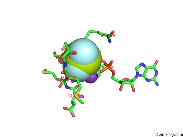
Fluorine binding site 1 out of 3 in 6jtg
Go back to
Fluorine binding site 1 out
of 3 in the Structural Insights Into G Domain Dimerization and Pathogenic Mutations of OPA1
Mono view

Stereo pair view
Mono view
Stereo pair view
A full contact list of Fluorine with other atoms in the F binding
site number 1 of Structural Insights Into G Domain Dimerization and Pathogenic Mutations of OPA1 within 5.0Å range:
|
Fluorine binding site 2 out of 3 in 6jtg
Go back to
Fluorine binding site 2 out
of 3 in the Structural Insights Into G Domain Dimerization and Pathogenic Mutations of OPA1

Mono view

Stereo pair view
Mono view
Stereo pair view
A full contact list of Fluorine with other atoms in the F binding
site number 2 of Structural Insights Into G Domain Dimerization and Pathogenic Mutations of OPA1 within 5.0Å range:
|
Fluorine binding site 3 out of 3 in 6jtg
Go back to
Fluorine binding site 3 out
of 3 in the Structural Insights Into G Domain Dimerization and Pathogenic Mutations of OPA1

Mono view

Stereo pair view
Mono view
Stereo pair view
A full contact list of Fluorine with other atoms in the F binding
site number 3 of Structural Insights Into G Domain Dimerization and Pathogenic Mutations of OPA1 within 5.0Å range:
|
Reference:
L.Yan,
J.Hu.
Structural Insights Into G Domain Dimerization and Pathogenic Mutations of OPA1 To Be Published.
Page generated: Tue Jul 15 12:42:55 2025
Last articles
Mg in 3A7RMg in 3A6P
Mg in 3A7E
Mg in 3A7D
Mg in 3A52
Mg in 3A64
Mg in 3A5T
Mg in 3A58
Mg in 3A5M
Mg in 3A5L