Fluorine »
PDB 6lub-6mii »
6m11 »
Fluorine in PDB 6m11: Crystal Structure of Rnase L in Complex with Sunitinib
Protein crystallography data
The structure of Crystal Structure of Rnase L in Complex with Sunitinib, PDB code: 6m11
was solved by
J.Tang,
H.Huang,
with X-Ray Crystallography technique. A brief refinement statistics is given in the table below:
| Resolution Low / High (Å) | 51.26 / 2.46 |
| Space group | P 21 21 21 |
| Cell size a, b, c (Å), α, β, γ (°) | 59.240, 111.000, 262.670, 90.00, 90.00, 90.00 |
| R / Rfree (%) | 22 / 28.5 |
Fluorine Binding Sites:
The binding sites of Fluorine atom in the Crystal Structure of Rnase L in Complex with Sunitinib
(pdb code 6m11). This binding sites where shown within
5.0 Angstroms radius around Fluorine atom.
In total 2 binding sites of Fluorine where determined in the Crystal Structure of Rnase L in Complex with Sunitinib, PDB code: 6m11:
Jump to Fluorine binding site number: 1; 2;
In total 2 binding sites of Fluorine where determined in the Crystal Structure of Rnase L in Complex with Sunitinib, PDB code: 6m11:
Jump to Fluorine binding site number: 1; 2;
Fluorine binding site 1 out of 2 in 6m11
Go back to
Fluorine binding site 1 out
of 2 in the Crystal Structure of Rnase L in Complex with Sunitinib
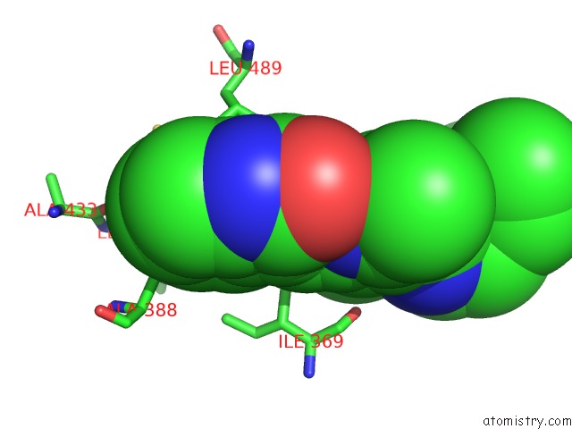
Mono view
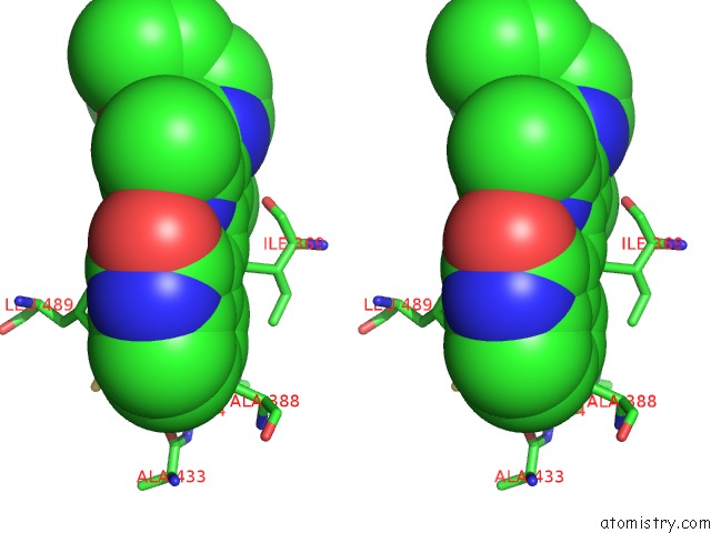
Stereo pair view
Mono view
Stereo pair view
A full contact list of Fluorine with other atoms in the F binding
site number 1 of Crystal Structure of Rnase L in Complex with Sunitinib within 5.0Å range:
|
Fluorine binding site 2 out of 2 in 6m11
Go back to
Fluorine binding site 2 out
of 2 in the Crystal Structure of Rnase L in Complex with Sunitinib

Mono view
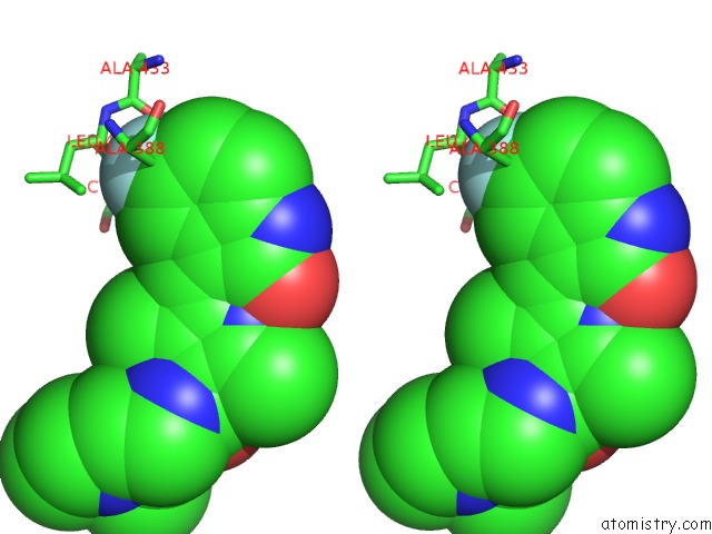
Stereo pair view
Mono view
Stereo pair view
A full contact list of Fluorine with other atoms in the F binding
site number 2 of Crystal Structure of Rnase L in Complex with Sunitinib within 5.0Å range:
|
Reference:
J.Tang,
Y.Wang,
H.Zhou,
Y.Ye,
M.Talukdar,
Z.Fu,
Z.Liu,
J.Li,
D.Neculai,
J.Gao,
H.Huang.
Sunitinib Inhibits Rnase L By Destabilizing Its Active Dimer Conformation. Biochem.J. V. 477 3387 2020.
ISSN: ESSN 1470-8728
PubMed: 32830849
DOI: 10.1042/BCJ20200260
Page generated: Tue Jul 15 13:03:45 2025
ISSN: ESSN 1470-8728
PubMed: 32830849
DOI: 10.1042/BCJ20200260
Last articles
Mg in 7BGIMg in 7BLX
Mg in 7BLZ
Mg in 7BOD
Mg in 7BNR
Mg in 7BNK
Mg in 7BMC
Mg in 7BM9
Mg in 7BM8
Mg in 7BM6