Fluorine »
PDB 6n4n-6ngq »
6nfz »
Fluorine in PDB 6nfz: Crystal Structure of Diphosphorylated HPK1 Kinase Domain in Complex with Sunitinib in the Active State.
Enzymatic activity of Crystal Structure of Diphosphorylated HPK1 Kinase Domain in Complex with Sunitinib in the Active State.
All present enzymatic activity of Crystal Structure of Diphosphorylated HPK1 Kinase Domain in Complex with Sunitinib in the Active State.:
2.7.11.1;
2.7.11.1;
Protein crystallography data
The structure of Crystal Structure of Diphosphorylated HPK1 Kinase Domain in Complex with Sunitinib in the Active State., PDB code: 6nfz
was solved by
E.Johnson,
M.Mctigue,
C.N.Cronin,
with X-Ray Crystallography technique. A brief refinement statistics is given in the table below:
| Resolution Low / High (Å) | 37.52 / 2.97 |
| Space group | H 3 2 |
| Cell size a, b, c (Å), α, β, γ (°) | 149.932, 149.932, 156.754, 90.00, 90.00, 120.00 |
| R / Rfree (%) | 20.7 / 25.1 |
Fluorine Binding Sites:
The binding sites of Fluorine atom in the Crystal Structure of Diphosphorylated HPK1 Kinase Domain in Complex with Sunitinib in the Active State.
(pdb code 6nfz). This binding sites where shown within
5.0 Angstroms radius around Fluorine atom.
In total 2 binding sites of Fluorine where determined in the Crystal Structure of Diphosphorylated HPK1 Kinase Domain in Complex with Sunitinib in the Active State., PDB code: 6nfz:
Jump to Fluorine binding site number: 1; 2;
In total 2 binding sites of Fluorine where determined in the Crystal Structure of Diphosphorylated HPK1 Kinase Domain in Complex with Sunitinib in the Active State., PDB code: 6nfz:
Jump to Fluorine binding site number: 1; 2;
Fluorine binding site 1 out of 2 in 6nfz
Go back to
Fluorine binding site 1 out
of 2 in the Crystal Structure of Diphosphorylated HPK1 Kinase Domain in Complex with Sunitinib in the Active State.
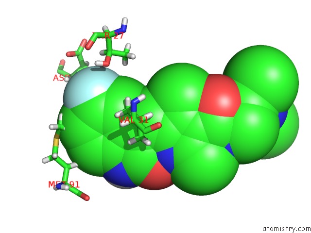
Mono view
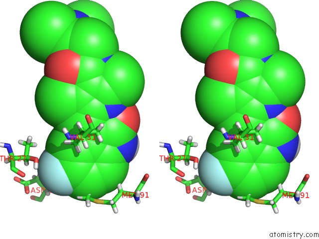
Stereo pair view
Mono view
Stereo pair view
A full contact list of Fluorine with other atoms in the F binding
site number 1 of Crystal Structure of Diphosphorylated HPK1 Kinase Domain in Complex with Sunitinib in the Active State. within 5.0Å range:
|
Fluorine binding site 2 out of 2 in 6nfz
Go back to
Fluorine binding site 2 out
of 2 in the Crystal Structure of Diphosphorylated HPK1 Kinase Domain in Complex with Sunitinib in the Active State.
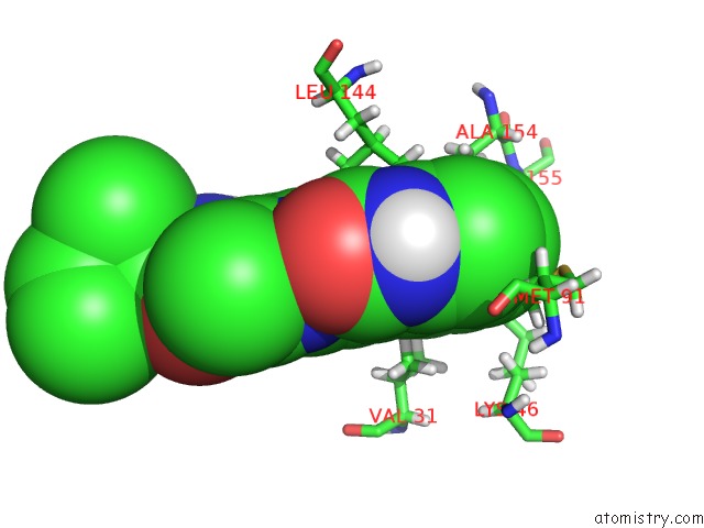
Mono view
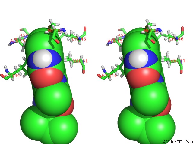
Stereo pair view
Mono view
Stereo pair view
A full contact list of Fluorine with other atoms in the F binding
site number 2 of Crystal Structure of Diphosphorylated HPK1 Kinase Domain in Complex with Sunitinib in the Active State. within 5.0Å range:
|
Reference:
E.Johnson,
M.Mctigue,
R.A.Gallego,
T.W.Johnson,
S.Timofeevski,
M.Maestre,
T.S.Fisher,
R.Kania,
S.Sawasdikosol,
S.Burakoff,
C.N.Cronin.
Multiple Conformational States of the HPK1 Kinase Domain in Complex with Sunitinib Reveal the Structural Changes Accompanying HPK1 Trans-Regulation. J.Biol.Chem. V. 294 9029 2019.
ISSN: ESSN 1083-351X
PubMed: 31018963
DOI: 10.1074/JBC.AC119.007466
Page generated: Tue Jul 15 13:22:31 2025
ISSN: ESSN 1083-351X
PubMed: 31018963
DOI: 10.1074/JBC.AC119.007466
Last articles
Mg in 4LCZMg in 4LF2
Mg in 4LF1
Mg in 4LEM
Mg in 4LCK
Mg in 4LE0
Mg in 4LDZ
Mg in 4LDT
Mg in 4LA7
Mg in 4LDJ