Fluorine »
PDB 6ony-6p3u »
6oyd »
Fluorine in PDB 6oyd: X-Ray Crystal Structure of Wild Type Hiv-1 Protease in Complex with Grl-004
Protein crystallography data
The structure of X-Ray Crystal Structure of Wild Type Hiv-1 Protease in Complex with Grl-004, PDB code: 6oyd
was solved by
H.Bulut,
S.I.Hattori,
H.Aoki-Ogata,
H.Hayashi,
M.Aoki,
A.K.Ghosh,
H.Mitsuya,
with X-Ray Crystallography technique. A brief refinement statistics is given in the table below:
| Resolution Low / High (Å) | 54.41 / 1.46 |
| Space group | P 61 2 2 |
| Cell size a, b, c (Å), α, β, γ (°) | 62.825, 62.825, 82.280, 90.00, 90.00, 120.00 |
| R / Rfree (%) | 21.8 / 25.7 |
Fluorine Binding Sites:
The binding sites of Fluorine atom in the X-Ray Crystal Structure of Wild Type Hiv-1 Protease in Complex with Grl-004
(pdb code 6oyd). This binding sites where shown within
5.0 Angstroms radius around Fluorine atom.
In total only one binding site of Fluorine was determined in the X-Ray Crystal Structure of Wild Type Hiv-1 Protease in Complex with Grl-004, PDB code: 6oyd:
In total only one binding site of Fluorine was determined in the X-Ray Crystal Structure of Wild Type Hiv-1 Protease in Complex with Grl-004, PDB code: 6oyd:
Fluorine binding site 1 out of 1 in 6oyd
Go back to
Fluorine binding site 1 out
of 1 in the X-Ray Crystal Structure of Wild Type Hiv-1 Protease in Complex with Grl-004
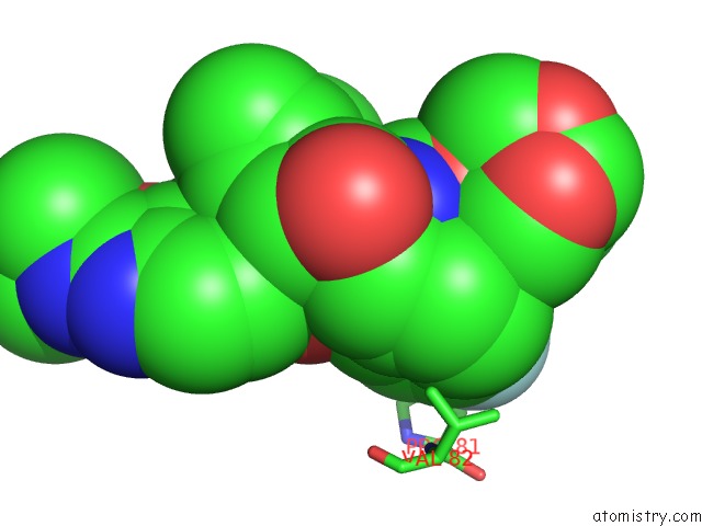
Mono view
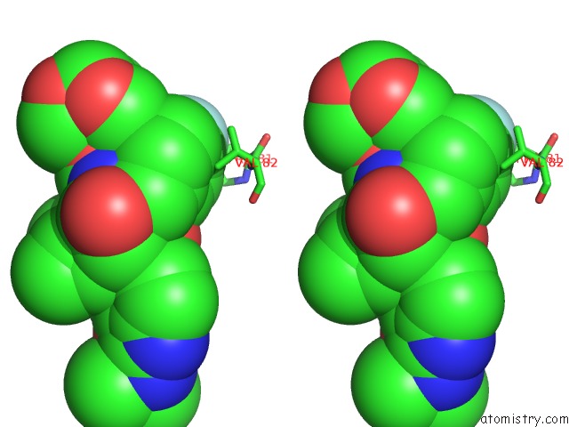
Stereo pair view
Mono view
Stereo pair view
A full contact list of Fluorine with other atoms in the F binding
site number 1 of X-Ray Crystal Structure of Wild Type Hiv-1 Protease in Complex with Grl-004 within 5.0Å range:
|
Reference:
H.Bulut,
S.I.Hattori,
H.Aoki-Ogata,
H.Hayashi,
M.Aoki,
A.K.Ghosh,
H.Mitsuya.
Novel Hiv-1 Protease Inhibitors, Grl-142 and Its Analogs, Markedly Adapt to the Structural Plasticity of Hiv-1 Protease and Exerts Extreme Potency with High Genetic Barrier To Be Published.
Page generated: Tue Jul 15 14:25:41 2025
Last articles
I in 4OWCI in 4P0D
I in 4OP3
I in 4OP2
I in 4OUC
I in 4OP1
I in 4OLH
I in 4OHP
I in 4OHO
I in 4OHM