Fluorine »
PDB 6r0h-6rkp »
6rj7 »
Fluorine in PDB 6rj7: Crystal Structure of the 19F Labelled Oxa-48
Enzymatic activity of Crystal Structure of the 19F Labelled Oxa-48
All present enzymatic activity of Crystal Structure of the 19F Labelled Oxa-48:
3.5.2.6;
3.5.2.6;
Protein crystallography data
The structure of Crystal Structure of the 19F Labelled Oxa-48, PDB code: 6rj7
was solved by
J.Brem,
C.Lohans,
C.Schofield,
with X-Ray Crystallography technique. A brief refinement statistics is given in the table below:
| Resolution Low / High (Å) | 66.40 / 1.73 |
| Space group | P 1 21 1 |
| Cell size a, b, c (Å), α, β, γ (°) | 64.360, 58.030, 66.430, 90.00, 91.49, 90.00 |
| R / Rfree (%) | 17.7 / 21 |
Other elements in 6rj7:
The structure of Crystal Structure of the 19F Labelled Oxa-48 also contains other interesting chemical elements:
| Calcium | (Ca) | 1 atom |
| Chlorine | (Cl) | 1 atom |
Fluorine Binding Sites:
The binding sites of Fluorine atom in the Crystal Structure of the 19F Labelled Oxa-48
(pdb code 6rj7). This binding sites where shown within
5.0 Angstroms radius around Fluorine atom.
In total 6 binding sites of Fluorine where determined in the Crystal Structure of the 19F Labelled Oxa-48, PDB code: 6rj7:
Jump to Fluorine binding site number: 1; 2; 3; 4; 5; 6;
In total 6 binding sites of Fluorine where determined in the Crystal Structure of the 19F Labelled Oxa-48, PDB code: 6rj7:
Jump to Fluorine binding site number: 1; 2; 3; 4; 5; 6;
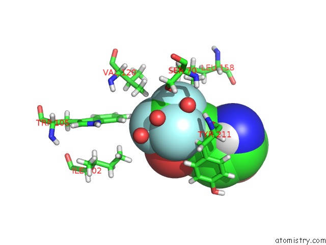
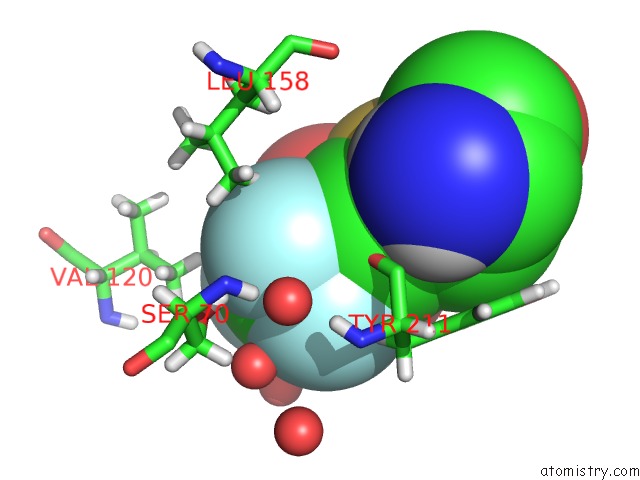
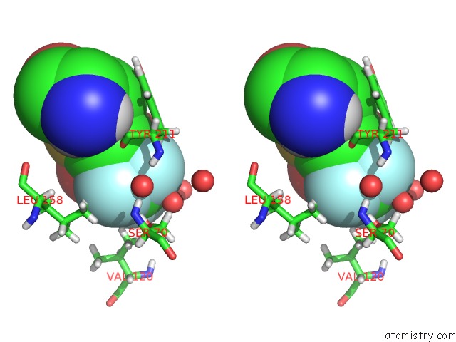
Fluorine binding site 1 out of 6 in 6rj7
Go back to
Fluorine binding site 1 out
of 6 in the Crystal Structure of the 19F Labelled Oxa-48
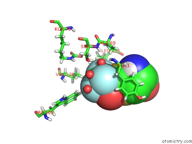
Mono view
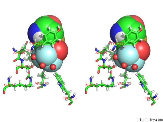
Stereo pair view
Mono view
Stereo pair view
A full contact list of Fluorine with other atoms in the F binding
site number 1 of Crystal Structure of the 19F Labelled Oxa-48 within 5.0Å range:
|
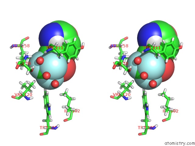
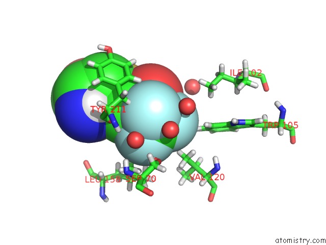
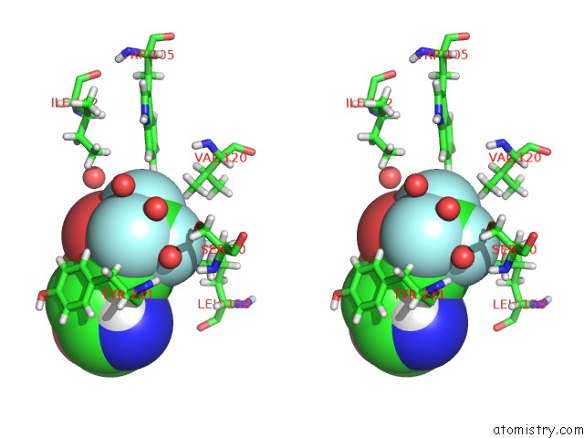
Fluorine binding site 2 out of 6 in 6rj7
Go back to
Fluorine binding site 2 out
of 6 in the Crystal Structure of the 19F Labelled Oxa-48
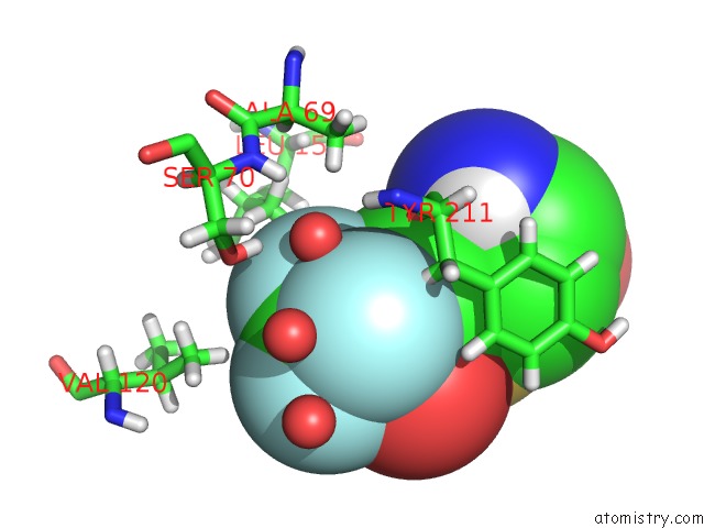
Mono view
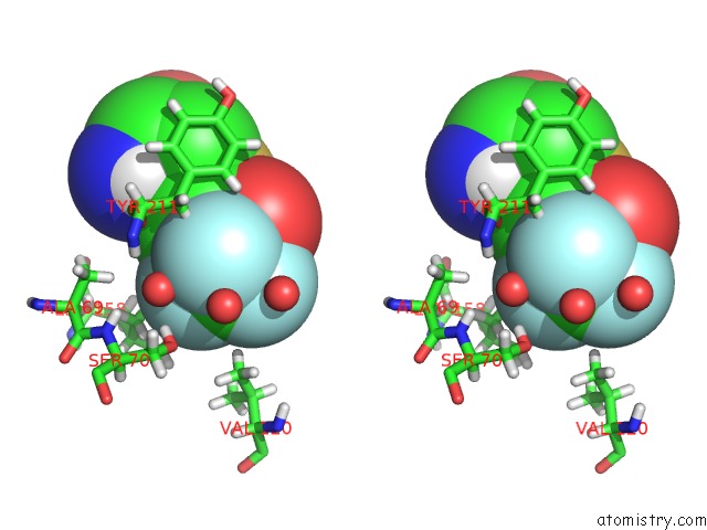
Stereo pair view
Mono view
Stereo pair view
A full contact list of Fluorine with other atoms in the F binding
site number 2 of Crystal Structure of the 19F Labelled Oxa-48 within 5.0Å range:
|
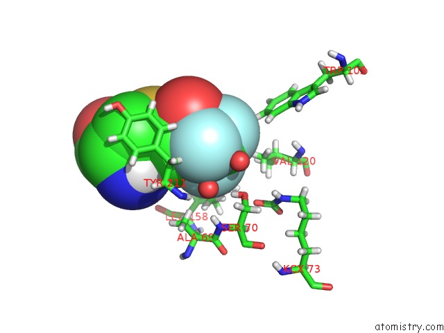
Fluorine binding site 3 out of 6 in 6rj7
Go back to
Fluorine binding site 3 out
of 6 in the Crystal Structure of the 19F Labelled Oxa-48
Mono view
Stereo pair view
Mono view
Stereo pair view
A full contact list of Fluorine with other atoms in the F binding
site number 3 of Crystal Structure of the 19F Labelled Oxa-48 within 5.0Å range:
|
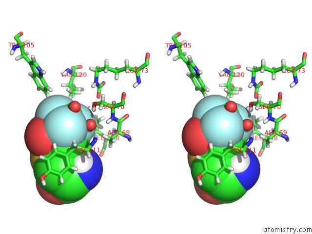
Fluorine binding site 4 out of 6 in 6rj7
Go back to
Fluorine binding site 4 out
of 6 in the Crystal Structure of the 19F Labelled Oxa-48
Mono view
Stereo pair view
Mono view
Stereo pair view
A full contact list of Fluorine with other atoms in the F binding
site number 4 of Crystal Structure of the 19F Labelled Oxa-48 within 5.0Å range:
|
Fluorine binding site 5 out of 6 in 6rj7
Go back to
Fluorine binding site 5 out
of 6 in the Crystal Structure of the 19F Labelled Oxa-48
Mono view
Stereo pair view
Mono view
Stereo pair view
A full contact list of Fluorine with other atoms in the F binding
site number 5 of Crystal Structure of the 19F Labelled Oxa-48 within 5.0Å range:
|
Fluorine binding site 6 out of 6 in 6rj7
Go back to
Fluorine binding site 6 out
of 6 in the Crystal Structure of the 19F Labelled Oxa-48
Mono view
Stereo pair view
Mono view
Stereo pair view
A full contact list of Fluorine with other atoms in the F binding
site number 6 of Crystal Structure of the 19F Labelled Oxa-48 within 5.0Å range:
|
Reference:
E.Van Groesen,
C.T.Lohans,
J.Brem,
K.M.J.Aertker,
T.D.W.Claridge,
C.J.Schofield.
19F uc(Nmr) Monitoring of Reversible Protein Post-Translational Modifications: Class D Beta-Lactamase Carbamylation and Inhibition. Chemistry V. 25 11837 2019.
ISSN: ISSN 0947-6539
PubMed: 31310409
DOI: 10.1002/CHEM.201902529
Page generated: Tue Jul 15 15:20:20 2025
ISSN: ISSN 0947-6539
PubMed: 31310409
DOI: 10.1002/CHEM.201902529
Last articles
Fe in 4QOMFe in 4QON
Fe in 4QOL
Fe in 4QO5
Fe in 4QM9
Fe in 4QDF
Fe in 4QMA
Fe in 4QM8
Fe in 4QLW
Fe in 4QI7