Fluorine »
PDB 6rzi-6slz »
6s1j »
Fluorine in PDB 6s1j: Crystal Structure of DYRK1A with Small Molecule Inhibitor
Enzymatic activity of Crystal Structure of DYRK1A with Small Molecule Inhibitor
All present enzymatic activity of Crystal Structure of DYRK1A with Small Molecule Inhibitor:
2.7.12.1;
2.7.12.1;
Protein crystallography data
The structure of Crystal Structure of DYRK1A with Small Molecule Inhibitor, PDB code: 6s1j
was solved by
F.J.Sorrell,
S.H.Henderson,
C.Redondo,
N.A.Burgess-Brown,
F.Von Delft,
C.H.Arrowsmith,
C.Bountra,
A.M.Edwards,
J.M.Elkins,
with X-Ray Crystallography technique. A brief refinement statistics is given in the table below:
| Resolution Low / High (Å) | 59.80 / 1.41 |
| Space group | C 1 2 1 |
| Cell size a, b, c (Å), α, β, γ (°) | 99.202, 69.671, 67.440, 90.00, 117.54, 90.00 |
| R / Rfree (%) | 15.5 / 17.6 |
Fluorine Binding Sites:
The binding sites of Fluorine atom in the Crystal Structure of DYRK1A with Small Molecule Inhibitor
(pdb code 6s1j). This binding sites where shown within
5.0 Angstroms radius around Fluorine atom.
In total only one binding site of Fluorine was determined in the Crystal Structure of DYRK1A with Small Molecule Inhibitor, PDB code: 6s1j:
In total only one binding site of Fluorine was determined in the Crystal Structure of DYRK1A with Small Molecule Inhibitor, PDB code: 6s1j:
Fluorine binding site 1 out of 1 in 6s1j
Go back to
Fluorine binding site 1 out
of 1 in the Crystal Structure of DYRK1A with Small Molecule Inhibitor

Mono view
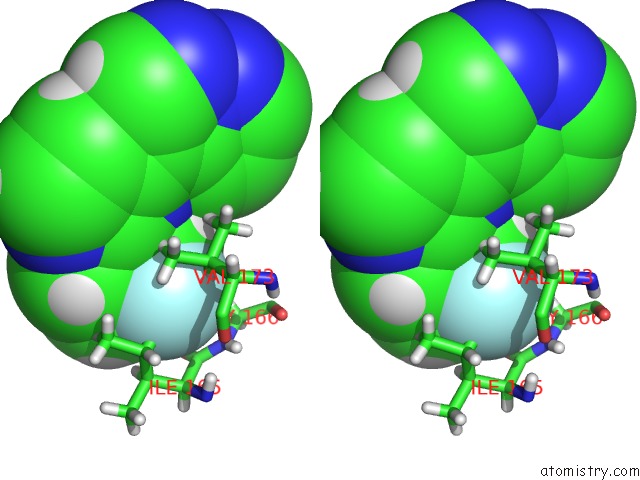
Stereo pair view
Mono view
Stereo pair view
A full contact list of Fluorine with other atoms in the F binding
site number 1 of Crystal Structure of DYRK1A with Small Molecule Inhibitor within 5.0Å range:
|
Reference:
F.J.Sorrell,
S.H.Henderson,
J.M.Elkins,
S.Ward.
Kinase Scaffold Repurposing in the Public Domain To Be Published.
Page generated: Tue Jul 15 15:34:59 2025
Last articles
Mg in 5G0RMg in 5G5V
Mg in 5G3T
Mg in 5G5T
Mg in 5G5S
Mg in 5G4A
Mg in 5G57
Mg in 5G50
Mg in 5G41
Mg in 5G3Z