Fluorine »
PDB 6sov-6taw »
6sq5 »
Fluorine in PDB 6sq5: Crystal Structure of M. Tuberculosis Inha in Complex with Nad+ and 3- [3-(Trifluoromethyl)Phenyl]Prop-2-Enoic Acid
Enzymatic activity of Crystal Structure of M. Tuberculosis Inha in Complex with Nad+ and 3- [3-(Trifluoromethyl)Phenyl]Prop-2-Enoic Acid
All present enzymatic activity of Crystal Structure of M. Tuberculosis Inha in Complex with Nad+ and 3- [3-(Trifluoromethyl)Phenyl]Prop-2-Enoic Acid:
1.3.1.9;
1.3.1.9;
Protein crystallography data
The structure of Crystal Structure of M. Tuberculosis Inha in Complex with Nad+ and 3- [3-(Trifluoromethyl)Phenyl]Prop-2-Enoic Acid, PDB code: 6sq5
was solved by
V.Mendes,
M.Sabbah,
A.G.Coyne,
C.Abell,
T.L.Blundell,
with X-Ray Crystallography technique. A brief refinement statistics is given in the table below:
| Resolution Low / High (Å) | 72.35 / 1.84 |
| Space group | P 62 2 2 |
| Cell size a, b, c (Å), α, β, γ (°) | 97.533, 97.533, 140.229, 90.00, 90.00, 120.00 |
| R / Rfree (%) | 16.3 / 18.2 |
Fluorine Binding Sites:
The binding sites of Fluorine atom in the Crystal Structure of M. Tuberculosis Inha in Complex with Nad+ and 3- [3-(Trifluoromethyl)Phenyl]Prop-2-Enoic Acid
(pdb code 6sq5). This binding sites where shown within
5.0 Angstroms radius around Fluorine atom.
In total 3 binding sites of Fluorine where determined in the Crystal Structure of M. Tuberculosis Inha in Complex with Nad+ and 3- [3-(Trifluoromethyl)Phenyl]Prop-2-Enoic Acid, PDB code: 6sq5:
Jump to Fluorine binding site number: 1; 2; 3;
In total 3 binding sites of Fluorine where determined in the Crystal Structure of M. Tuberculosis Inha in Complex with Nad+ and 3- [3-(Trifluoromethyl)Phenyl]Prop-2-Enoic Acid, PDB code: 6sq5:
Jump to Fluorine binding site number: 1; 2; 3;
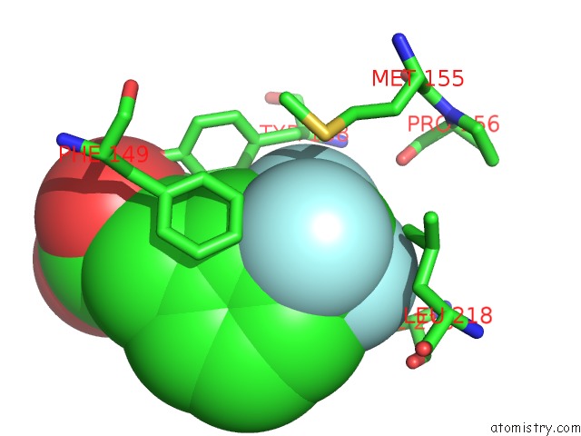
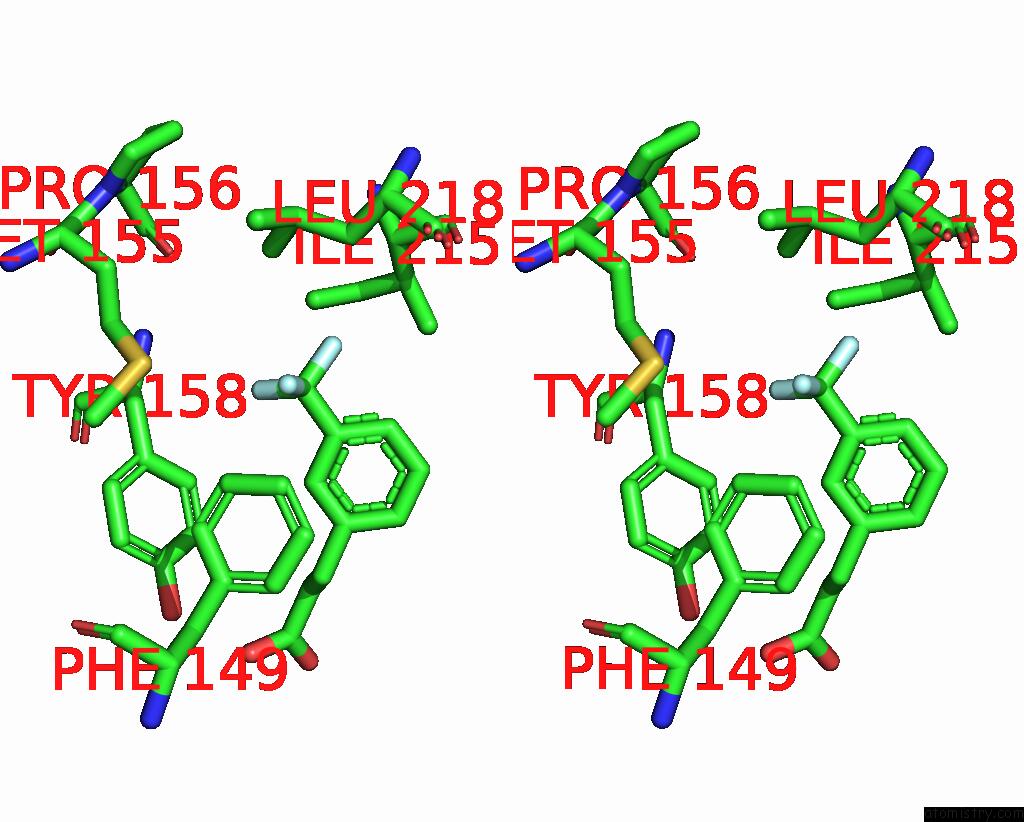
Fluorine binding site 1 out of 3 in 6sq5
Go back to
Fluorine binding site 1 out
of 3 in the Crystal Structure of M. Tuberculosis Inha in Complex with Nad+ and 3- [3-(Trifluoromethyl)Phenyl]Prop-2-Enoic Acid
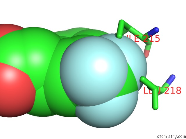
Mono view
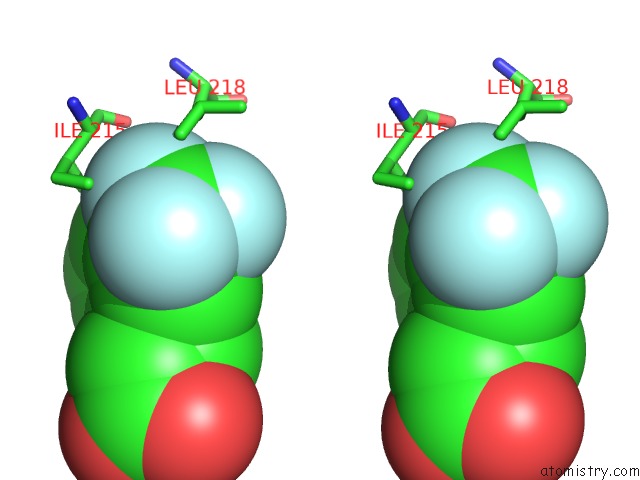
Stereo pair view
Mono view
Stereo pair view
A full contact list of Fluorine with other atoms in the F binding
site number 1 of Crystal Structure of M. Tuberculosis Inha in Complex with Nad+ and 3- [3-(Trifluoromethyl)Phenyl]Prop-2-Enoic Acid within 5.0Å range:
|
Fluorine binding site 2 out of 3 in 6sq5
Go back to
Fluorine binding site 2 out
of 3 in the Crystal Structure of M. Tuberculosis Inha in Complex with Nad+ and 3- [3-(Trifluoromethyl)Phenyl]Prop-2-Enoic Acid
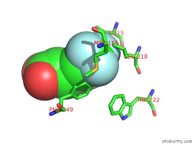
Mono view
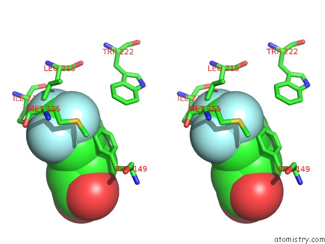
Stereo pair view
Mono view
Stereo pair view
A full contact list of Fluorine with other atoms in the F binding
site number 2 of Crystal Structure of M. Tuberculosis Inha in Complex with Nad+ and 3- [3-(Trifluoromethyl)Phenyl]Prop-2-Enoic Acid within 5.0Å range:
|
Fluorine binding site 3 out of 3 in 6sq5
Go back to
Fluorine binding site 3 out
of 3 in the Crystal Structure of M. Tuberculosis Inha in Complex with Nad+ and 3- [3-(Trifluoromethyl)Phenyl]Prop-2-Enoic Acid
Mono view
Stereo pair view
Mono view
Stereo pair view
A full contact list of Fluorine with other atoms in the F binding
site number 3 of Crystal Structure of M. Tuberculosis Inha in Complex with Nad+ and 3- [3-(Trifluoromethyl)Phenyl]Prop-2-Enoic Acid within 5.0Å range:
|
Reference:
M.Sabbah,
V.Mendes,
R.G.Vistal,
D.M.Dias,
M.Zahorszka,
K.Mikusova,
J.Kordulakova,
A.G.Coyne,
T.L.Blundell,
C.Abell.
Fragment-Based Design of Mycobacterium Tuberculosis Inha Inhibitors. J.Med.Chem. 2020.
ISSN: ISSN 0022-2623
PubMed: 32240584
DOI: 10.1021/ACS.JMEDCHEM.0C00007
Page generated: Tue Jul 15 15:45:19 2025
ISSN: ISSN 0022-2623
PubMed: 32240584
DOI: 10.1021/ACS.JMEDCHEM.0C00007
Last articles
Fe in 8PYAFe in 8PY8
Fe in 8PY7
Fe in 8PY6
Fe in 8PY9
Fe in 8PVM
Fe in 8PWY
Fe in 8PXQ
Fe in 8PY5
Fe in 8PWS