Fluorine »
PDB 6tvl-6ugr »
6udm »
Fluorine in PDB 6udm: Structure of Human Cytochrome P450 1A1 with Duocarmycin Prodrug (S) Ict-2726
Enzymatic activity of Structure of Human Cytochrome P450 1A1 with Duocarmycin Prodrug (S) Ict-2726
All present enzymatic activity of Structure of Human Cytochrome P450 1A1 with Duocarmycin Prodrug (S) Ict-2726:
1.14.14.1; 4.2.1.152;
1.14.14.1; 4.2.1.152;
Protein crystallography data
The structure of Structure of Human Cytochrome P450 1A1 with Duocarmycin Prodrug (S) Ict-2726, PDB code: 6udm
was solved by
A.G.Bart,
E.E.Scott,
with X-Ray Crystallography technique. A brief refinement statistics is given in the table below:
| Resolution Low / High (Å) | 39.64 / 3.08 |
| Space group | P 31 2 1 |
| Cell size a, b, c (Å), α, β, γ (°) | 241.281, 241.281, 125.298, 90.00, 90.00, 120.00 |
| R / Rfree (%) | 20.9 / 22.1 |
Other elements in 6udm:
The structure of Structure of Human Cytochrome P450 1A1 with Duocarmycin Prodrug (S) Ict-2726 also contains other interesting chemical elements:
| Iron | (Fe) | 4 atoms |
| Chlorine | (Cl) | 4 atoms |
Fluorine Binding Sites:
The binding sites of Fluorine atom in the Structure of Human Cytochrome P450 1A1 with Duocarmycin Prodrug (S) Ict-2726
(pdb code 6udm). This binding sites where shown within
5.0 Angstroms radius around Fluorine atom.
In total 4 binding sites of Fluorine where determined in the Structure of Human Cytochrome P450 1A1 with Duocarmycin Prodrug (S) Ict-2726, PDB code: 6udm:
Jump to Fluorine binding site number: 1; 2; 3; 4;
In total 4 binding sites of Fluorine where determined in the Structure of Human Cytochrome P450 1A1 with Duocarmycin Prodrug (S) Ict-2726, PDB code: 6udm:
Jump to Fluorine binding site number: 1; 2; 3; 4;
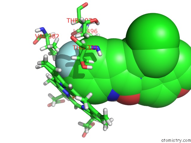
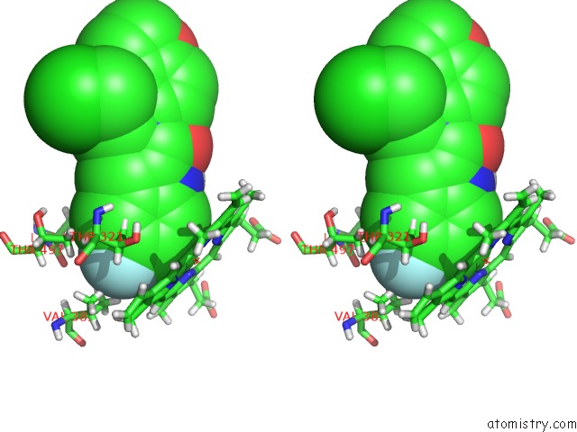
Fluorine binding site 1 out of 4 in 6udm
Go back to
Fluorine binding site 1 out
of 4 in the Structure of Human Cytochrome P450 1A1 with Duocarmycin Prodrug (S) Ict-2726
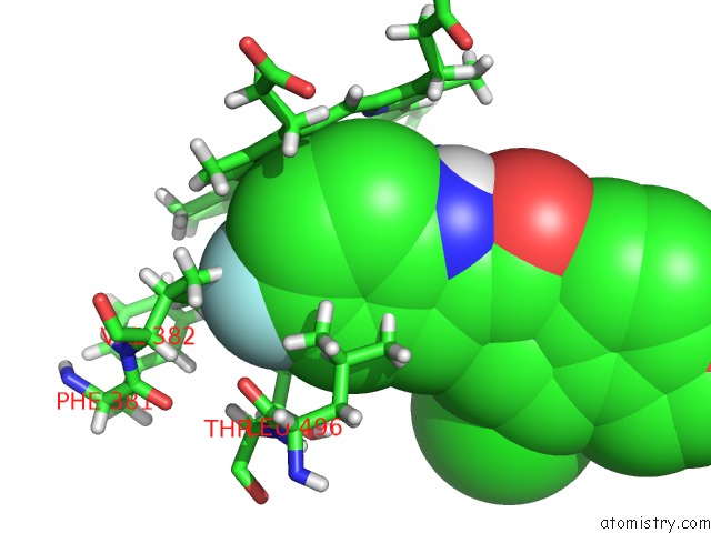
Mono view
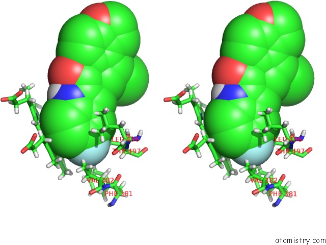
Stereo pair view
Mono view
Stereo pair view
A full contact list of Fluorine with other atoms in the F binding
site number 1 of Structure of Human Cytochrome P450 1A1 with Duocarmycin Prodrug (S) Ict-2726 within 5.0Å range:
|
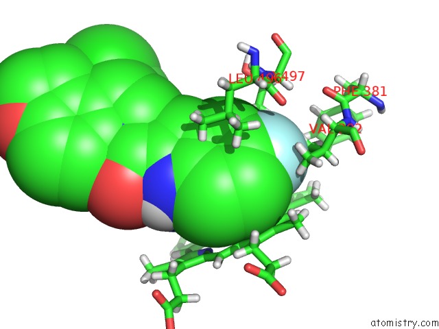
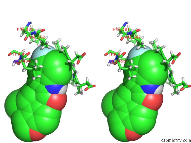
Fluorine binding site 2 out of 4 in 6udm
Go back to
Fluorine binding site 2 out
of 4 in the Structure of Human Cytochrome P450 1A1 with Duocarmycin Prodrug (S) Ict-2726
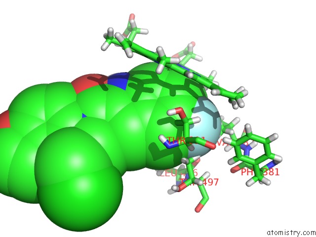
Mono view
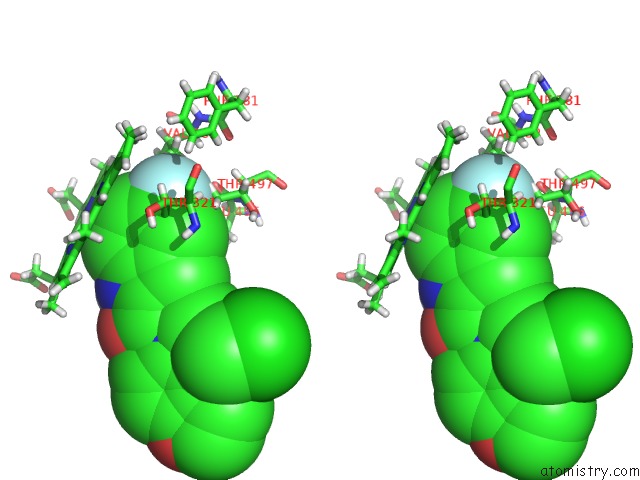
Stereo pair view
Mono view
Stereo pair view
A full contact list of Fluorine with other atoms in the F binding
site number 2 of Structure of Human Cytochrome P450 1A1 with Duocarmycin Prodrug (S) Ict-2726 within 5.0Å range:
|
Fluorine binding site 3 out of 4 in 6udm
Go back to
Fluorine binding site 3 out
of 4 in the Structure of Human Cytochrome P450 1A1 with Duocarmycin Prodrug (S) Ict-2726
Mono view
Stereo pair view
Mono view
Stereo pair view
A full contact list of Fluorine with other atoms in the F binding
site number 3 of Structure of Human Cytochrome P450 1A1 with Duocarmycin Prodrug (S) Ict-2726 within 5.0Å range:
|
Fluorine binding site 4 out of 4 in 6udm
Go back to
Fluorine binding site 4 out
of 4 in the Structure of Human Cytochrome P450 1A1 with Duocarmycin Prodrug (S) Ict-2726
Mono view
Stereo pair view
Mono view
Stereo pair view
A full contact list of Fluorine with other atoms in the F binding
site number 4 of Structure of Human Cytochrome P450 1A1 with Duocarmycin Prodrug (S) Ict-2726 within 5.0Å range:
|
Reference:
A.G.Bart,
E.E.Scott.
Designed Duocarmycin Prodrugs For Human Cytochrome P450 1A1 and 2W1 To Be Published.
Page generated: Tue Jul 15 16:15:52 2025
Last articles
K in 7QK5K in 7QIX
K in 7QNO
K in 7QIY
K in 7Q3X
K in 7QDN
K in 7QF6
K in 7Q0G
K in 7Q1C
K in 7Q1B