Fluorine »
PDB 6ugz-6v34 »
6uuo »
Fluorine in PDB 6uuo: Crystal Structure of Braf Kinase Domain Bound to the Protac P4B
Enzymatic activity of Crystal Structure of Braf Kinase Domain Bound to the Protac P4B
All present enzymatic activity of Crystal Structure of Braf Kinase Domain Bound to the Protac P4B:
2.7.11.1;
2.7.11.1;
Protein crystallography data
The structure of Crystal Structure of Braf Kinase Domain Bound to the Protac P4B, PDB code: 6uuo
was solved by
P.Maisonneuve,
G.Posternak,
I.Kurinov,
F.Sicheri,
with X-Ray Crystallography technique. A brief refinement statistics is given in the table below:
| Resolution Low / High (Å) | 54.95 / 3.29 |
| Space group | P 21 21 21 |
| Cell size a, b, c (Å), α, β, γ (°) | 52.059, 104.244, 109.904, 90.00, 90.00, 90.00 |
| R / Rfree (%) | 26.3 / 28.8 |
Fluorine Binding Sites:
The binding sites of Fluorine atom in the Crystal Structure of Braf Kinase Domain Bound to the Protac P4B
(pdb code 6uuo). This binding sites where shown within
5.0 Angstroms radius around Fluorine atom.
In total 4 binding sites of Fluorine where determined in the Crystal Structure of Braf Kinase Domain Bound to the Protac P4B, PDB code: 6uuo:
Jump to Fluorine binding site number: 1; 2; 3; 4;
In total 4 binding sites of Fluorine where determined in the Crystal Structure of Braf Kinase Domain Bound to the Protac P4B, PDB code: 6uuo:
Jump to Fluorine binding site number: 1; 2; 3; 4;
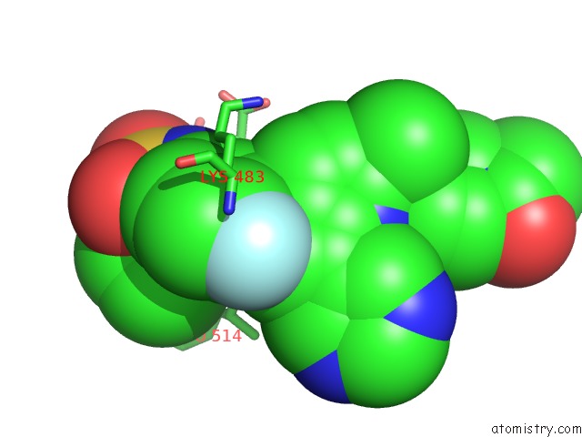
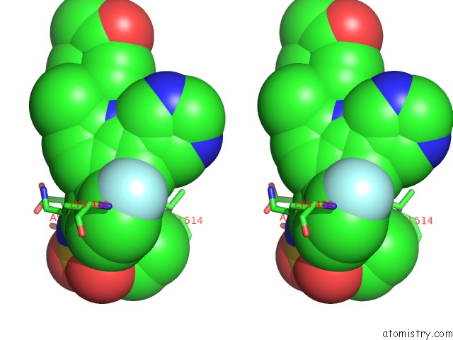
Fluorine binding site 1 out of 4 in 6uuo
Go back to
Fluorine binding site 1 out
of 4 in the Crystal Structure of Braf Kinase Domain Bound to the Protac P4B
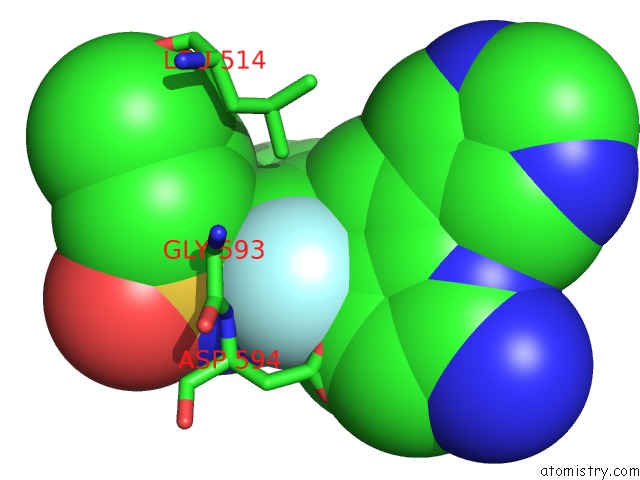
Mono view
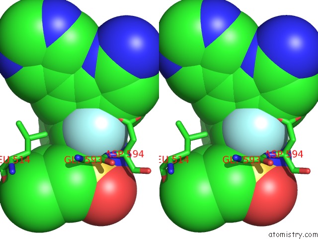
Stereo pair view
Mono view
Stereo pair view
A full contact list of Fluorine with other atoms in the F binding
site number 1 of Crystal Structure of Braf Kinase Domain Bound to the Protac P4B within 5.0Å range:
|
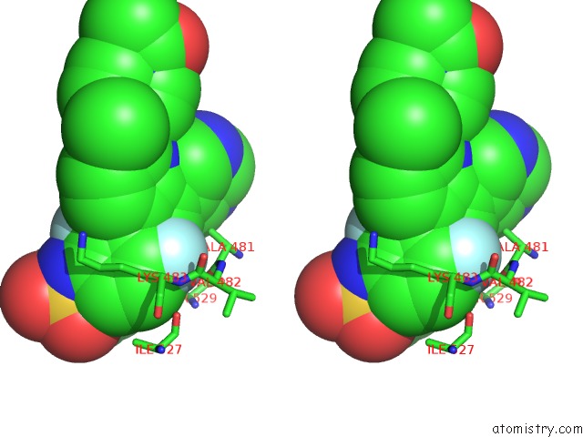
Fluorine binding site 2 out of 4 in 6uuo
Go back to
Fluorine binding site 2 out
of 4 in the Crystal Structure of Braf Kinase Domain Bound to the Protac P4B
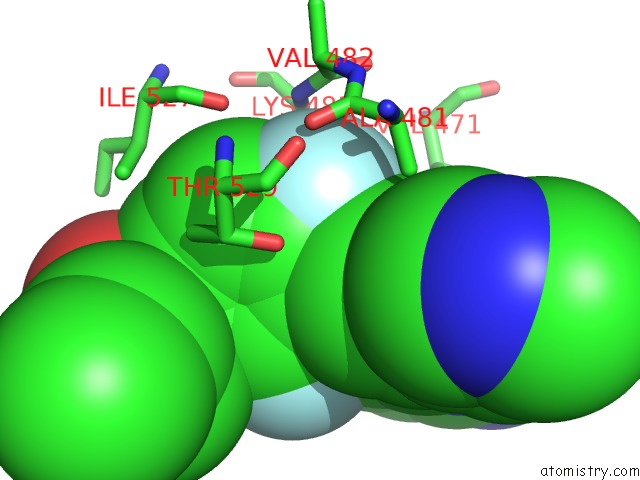
Mono view
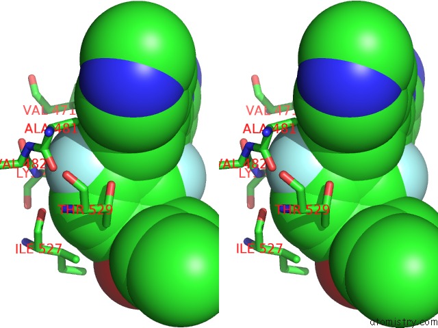
Stereo pair view
Mono view
Stereo pair view
A full contact list of Fluorine with other atoms in the F binding
site number 2 of Crystal Structure of Braf Kinase Domain Bound to the Protac P4B within 5.0Å range:
|
Fluorine binding site 3 out of 4 in 6uuo
Go back to
Fluorine binding site 3 out
of 4 in the Crystal Structure of Braf Kinase Domain Bound to the Protac P4B
Mono view
Stereo pair view
Mono view
Stereo pair view
A full contact list of Fluorine with other atoms in the F binding
site number 3 of Crystal Structure of Braf Kinase Domain Bound to the Protac P4B within 5.0Å range:
|
Fluorine binding site 4 out of 4 in 6uuo
Go back to
Fluorine binding site 4 out
of 4 in the Crystal Structure of Braf Kinase Domain Bound to the Protac P4B

Mono view
Stereo pair view
Mono view
Stereo pair view
A full contact list of Fluorine with other atoms in the F binding
site number 4 of Crystal Structure of Braf Kinase Domain Bound to the Protac P4B within 5.0Å range:
|
Reference:
P.Maisonneuve,
X.Tang,
G.Posternak,
T.Jin,
Z.Yin,
C.Colwill,
B.Larsen,
C.Wong,
S.Maier,
A.C.Gingras,
R.Batey,
M.Taipale,
A.Aman,
M.Prakesch,
G.Poda,
D.Uehling,
R.Al-Awar,
H.Lavoie,
I.Kurinov,
M.Therrien,
F.Sicheri.
Functional Characterization of A Protac Directed Against BRAFV600E To Be Published.
Page generated: Tue Jul 15 16:20:37 2025
Last articles
Na in 2XGENa in 2XDJ
Na in 2XCJ
Na in 2XC6
Na in 2XDC
Na in 2XC5
Na in 2XC4
Na in 2X8J
Na in 2XC0
Na in 2XBY