Fluorine »
PDB 6ugz-6v34 »
6uy4 »
Fluorine in PDB 6uy4: Crystal Structure of Dihydroorotate Dehydrogenase From Schistosoma Mansoni
Enzymatic activity of Crystal Structure of Dihydroorotate Dehydrogenase From Schistosoma Mansoni
All present enzymatic activity of Crystal Structure of Dihydroorotate Dehydrogenase From Schistosoma Mansoni:
1.3.98.1;
1.3.98.1;
Protein crystallography data
The structure of Crystal Structure of Dihydroorotate Dehydrogenase From Schistosoma Mansoni, PDB code: 6uy4
was solved by
R.M.Mori,
L.C.C.Zapata,
M.C.Nonato,
with X-Ray Crystallography technique. A brief refinement statistics is given in the table below:
| Resolution Low / High (Å) | 47.00 / 2.80 |
| Space group | I 4 3 2 |
| Cell size a, b, c (Å), α, β, γ (°) | 187.981, 187.981, 187.981, 90.00, 90.00, 90.00 |
| R / Rfree (%) | 21.6 / 24.6 |
Fluorine Binding Sites:
The binding sites of Fluorine atom in the Crystal Structure of Dihydroorotate Dehydrogenase From Schistosoma Mansoni
(pdb code 6uy4). This binding sites where shown within
5.0 Angstroms radius around Fluorine atom.
In total only one binding site of Fluorine was determined in the Crystal Structure of Dihydroorotate Dehydrogenase From Schistosoma Mansoni, PDB code: 6uy4:
In total only one binding site of Fluorine was determined in the Crystal Structure of Dihydroorotate Dehydrogenase From Schistosoma Mansoni, PDB code: 6uy4:
Fluorine binding site 1 out of 1 in 6uy4
Go back to
Fluorine binding site 1 out
of 1 in the Crystal Structure of Dihydroorotate Dehydrogenase From Schistosoma Mansoni
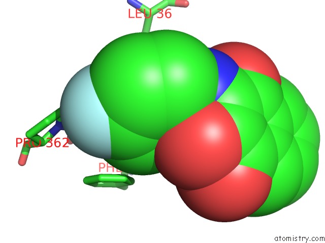
Mono view
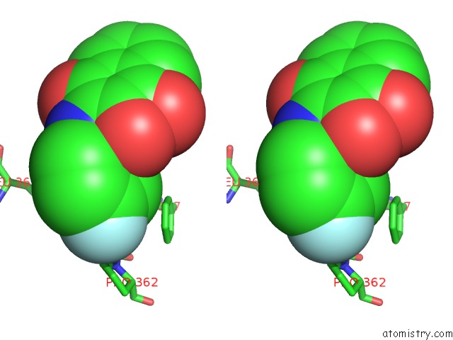
Stereo pair view
Mono view
Stereo pair view
A full contact list of Fluorine with other atoms in the F binding
site number 1 of Crystal Structure of Dihydroorotate Dehydrogenase From Schistosoma Mansoni within 5.0Å range:
|
Reference:
R.M.Mori,
M.A.A.Aleixo,
L.C.C.Zapata,
F.A.Calil,
F.D.S.Emery,
M.C.Nonato.
Structural Basis For the Function and Inhibition of Dihydroorotate Dehydrogenase From Schistosoma Mansoni. Febs J. 2020.
ISSN: ISSN 1742-464X
PubMed: 32428996
DOI: 10.1111/FEBS.15367
Page generated: Tue Jul 15 16:23:17 2025
ISSN: ISSN 1742-464X
PubMed: 32428996
DOI: 10.1111/FEBS.15367
Last articles
Na in 4K3INa in 4K7T
Na in 4K6V
Na in 4K6A
Na in 4K10
Na in 4JZZ
Na in 4K0W
Na in 4K28
Na in 4K19
Na in 4K0D