Fluorine »
PDB 7bjd-7cky »
7c7o »
Fluorine in PDB 7c7o: Crystal Structure of E.Coli Dna Gyrase B in Complex with 6-Fluoro-8- (Methylamino)-2-Oxo-1,2-Dihydroquinoline Derivative
Enzymatic activity of Crystal Structure of E.Coli Dna Gyrase B in Complex with 6-Fluoro-8- (Methylamino)-2-Oxo-1,2-Dihydroquinoline Derivative
All present enzymatic activity of Crystal Structure of E.Coli Dna Gyrase B in Complex with 6-Fluoro-8- (Methylamino)-2-Oxo-1,2-Dihydroquinoline Derivative:
5.6.2.2;
5.6.2.2;
Protein crystallography data
The structure of Crystal Structure of E.Coli Dna Gyrase B in Complex with 6-Fluoro-8- (Methylamino)-2-Oxo-1,2-Dihydroquinoline Derivative, PDB code: 7c7o
was solved by
M.Kamitani,
M.Mima,
T.Takeuchi,
F.Ushiyama,
with X-Ray Crystallography technique. A brief refinement statistics is given in the table below:
| Resolution Low / High (Å) | 17.61 / 1.80 |
| Space group | P 21 21 21 |
| Cell size a, b, c (Å), α, β, γ (°) | 41.251, 67.585, 67.913, 90.00, 90.00, 90.00 |
| R / Rfree (%) | 17 / 22.7 |
Fluorine Binding Sites:
The binding sites of Fluorine atom in the Crystal Structure of E.Coli Dna Gyrase B in Complex with 6-Fluoro-8- (Methylamino)-2-Oxo-1,2-Dihydroquinoline Derivative
(pdb code 7c7o). This binding sites where shown within
5.0 Angstroms radius around Fluorine atom.
In total only one binding site of Fluorine was determined in the Crystal Structure of E.Coli Dna Gyrase B in Complex with 6-Fluoro-8- (Methylamino)-2-Oxo-1,2-Dihydroquinoline Derivative, PDB code: 7c7o:
In total only one binding site of Fluorine was determined in the Crystal Structure of E.Coli Dna Gyrase B in Complex with 6-Fluoro-8- (Methylamino)-2-Oxo-1,2-Dihydroquinoline Derivative, PDB code: 7c7o:
Fluorine binding site 1 out of 1 in 7c7o
Go back to
Fluorine binding site 1 out
of 1 in the Crystal Structure of E.Coli Dna Gyrase B in Complex with 6-Fluoro-8- (Methylamino)-2-Oxo-1,2-Dihydroquinoline Derivative
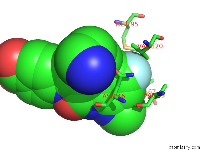
Mono view
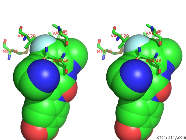
Stereo pair view
Mono view
Stereo pair view
A full contact list of Fluorine with other atoms in the F binding
site number 1 of Crystal Structure of E.Coli Dna Gyrase B in Complex with 6-Fluoro-8- (Methylamino)-2-Oxo-1,2-Dihydroquinoline Derivative within 5.0Å range:
|
Reference:
F.Ushiyama,
H.Amada,
Y.Mihara,
T.Takeuchi,
N.Tanaka-Yamamoto,
M.Mima,
M.Kamitani,
R.Wada,
Y.Tamura,
M.Endo,
A.Masuko,
I.Takata,
K.Hitaka,
H.Sugiyama,
N.Ohtake.
Lead Optimization of 8-(Methylamino)-2-Oxo-1,2-Dihydroquinolines As Bacterial Type II Topoisomerase Inhibitors. Bioorg.Med.Chem. V. 28 15776 2020.
ISSN: ESSN 1464-3391
PubMed: 33032189
DOI: 10.1016/J.BMC.2020.115776
Page generated: Tue Jul 15 18:53:35 2025
ISSN: ESSN 1464-3391
PubMed: 33032189
DOI: 10.1016/J.BMC.2020.115776
Last articles
Fe in 2YXOFe in 2YRS
Fe in 2YXC
Fe in 2YNM
Fe in 2YVJ
Fe in 2YP1
Fe in 2YU2
Fe in 2YU1
Fe in 2YQB
Fe in 2YOO