Fluorine »
PDB 7dcz-7e5i »
7dso »
Fluorine in PDB 7dso: Anthranilate Phosphoribosyltransferase From Saccharomyces Cerevisiae in Complex with 4-Fluoroanthranilate
Enzymatic activity of Anthranilate Phosphoribosyltransferase From Saccharomyces Cerevisiae in Complex with 4-Fluoroanthranilate
All present enzymatic activity of Anthranilate Phosphoribosyltransferase From Saccharomyces Cerevisiae in Complex with 4-Fluoroanthranilate:
2.4.2.18;
2.4.2.18;
Protein crystallography data
The structure of Anthranilate Phosphoribosyltransferase From Saccharomyces Cerevisiae in Complex with 4-Fluoroanthranilate, PDB code: 7dso
was solved by
X.Wu,
with X-Ray Crystallography technique. A brief refinement statistics is given in the table below:
| Resolution Low / High (Å) | 46.92 / 2.34 |
| Space group | P 21 21 21 |
| Cell size a, b, c (Å), α, β, γ (°) | 80.02, 87.914, 110.834, 90, 90, 90 |
| R / Rfree (%) | 17.7 / 23.9 |
Fluorine Binding Sites:
The binding sites of Fluorine atom in the Anthranilate Phosphoribosyltransferase From Saccharomyces Cerevisiae in Complex with 4-Fluoroanthranilate
(pdb code 7dso). This binding sites where shown within
5.0 Angstroms radius around Fluorine atom.
In total only one binding site of Fluorine was determined in the Anthranilate Phosphoribosyltransferase From Saccharomyces Cerevisiae in Complex with 4-Fluoroanthranilate, PDB code: 7dso:
In total only one binding site of Fluorine was determined in the Anthranilate Phosphoribosyltransferase From Saccharomyces Cerevisiae in Complex with 4-Fluoroanthranilate, PDB code: 7dso:
Fluorine binding site 1 out of 1 in 7dso
Go back to
Fluorine binding site 1 out
of 1 in the Anthranilate Phosphoribosyltransferase From Saccharomyces Cerevisiae in Complex with 4-Fluoroanthranilate
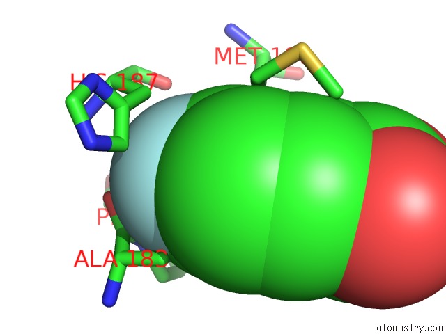
Mono view
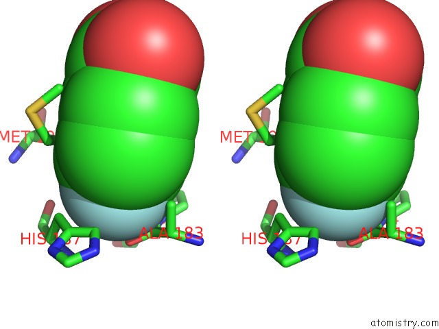
Stereo pair view
Mono view
Stereo pair view
A full contact list of Fluorine with other atoms in the F binding
site number 1 of Anthranilate Phosphoribosyltransferase From Saccharomyces Cerevisiae in Complex with 4-Fluoroanthranilate within 5.0Å range:
|
Reference:
X.Wu,
M.Zhang,
Z.Kuang,
J.Yue,
L.Xue,
M.Zhu,
Z.Zhu,
M.H.Khan,
L.Niu.
Crystal Structures of Anthranilate Phosphoribosyltransferase From Saccharomyces Cerevisiae. Acta Crystallogr.,Sect.F V. 77 61 2021.
ISSN: ESSN 2053-230X
PubMed: 33682790
DOI: 10.1107/S2053230X21001989
Page generated: Tue Jul 15 19:10:45 2025
ISSN: ESSN 2053-230X
PubMed: 33682790
DOI: 10.1107/S2053230X21001989
Last articles
Mg in 4W5OMg in 4W5J
Mg in 4W5N
Mg in 4V2I
Mg in 4V3R
Mg in 4V26
Mg in 4V2G
Mg in 4V1T
Mg in 4V25
Mg in 4V1V