Fluorine »
PDB 7f97-7fmn »
7feu »
Fluorine in PDB 7feu: The 0.95 Angstrom X-Ray Structure of the Human Heart Fatty Acid- Binding Protein Complexed with Perfluorononanoic Acid
Protein crystallography data
The structure of The 0.95 Angstrom X-Ray Structure of the Human Heart Fatty Acid- Binding Protein Complexed with Perfluorononanoic Acid, PDB code: 7feu
was solved by
S.Sugiyama,
K.Kakinouchi,
T.Hara,
R.Nakano,
S.Matsuoka,
H.Tsuchikawa,
M.Sonoyama,
Y.Inoue,
F.Hayashi,
M.Murata,
with X-Ray Crystallography technique. A brief refinement statistics is given in the table below:
| Resolution Low / High (Å) | 29.39 / 0.95 |
| Space group | P 21 21 21 |
| Cell size a, b, c (Å), α, β, γ (°) | 54.721, 69.615, 33.832, 90, 90, 90 |
| R / Rfree (%) | 11.7 / 13.8 |
Fluorine Binding Sites:
Pages:
>>> Page 1 <<< Page 2, Binding sites: 11 - 17;Binding sites:
The binding sites of Fluorine atom in the The 0.95 Angstrom X-Ray Structure of the Human Heart Fatty Acid- Binding Protein Complexed with Perfluorononanoic Acid (pdb code 7feu). This binding sites where shown within 5.0 Angstroms radius around Fluorine atom.In total 17 binding sites of Fluorine where determined in the The 0.95 Angstrom X-Ray Structure of the Human Heart Fatty Acid- Binding Protein Complexed with Perfluorononanoic Acid, PDB code: 7feu:
Jump to Fluorine binding site number: 1; 2; 3; 4; 5; 6; 7; 8; 9; 10;
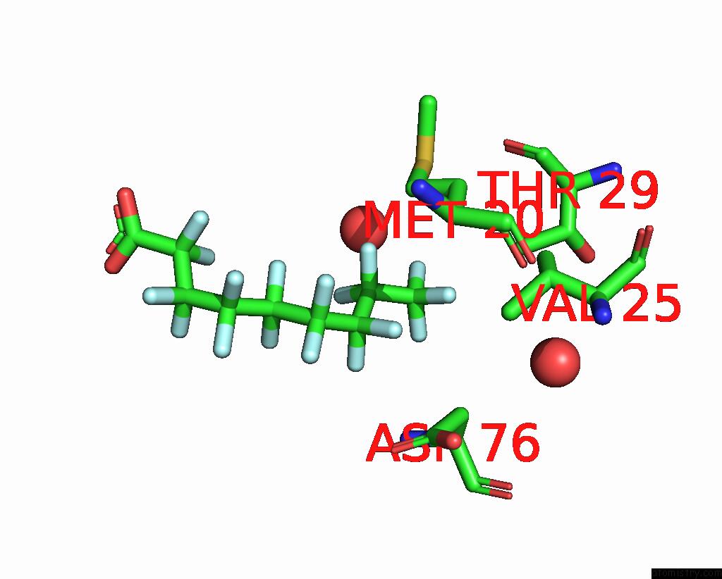
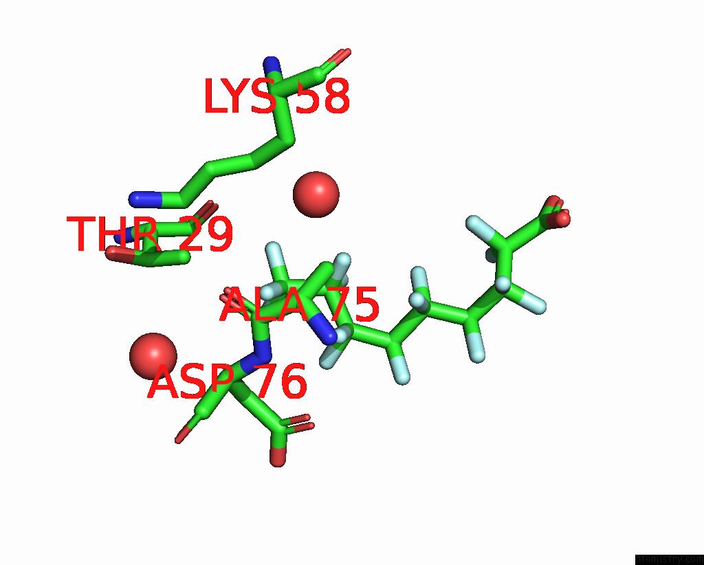
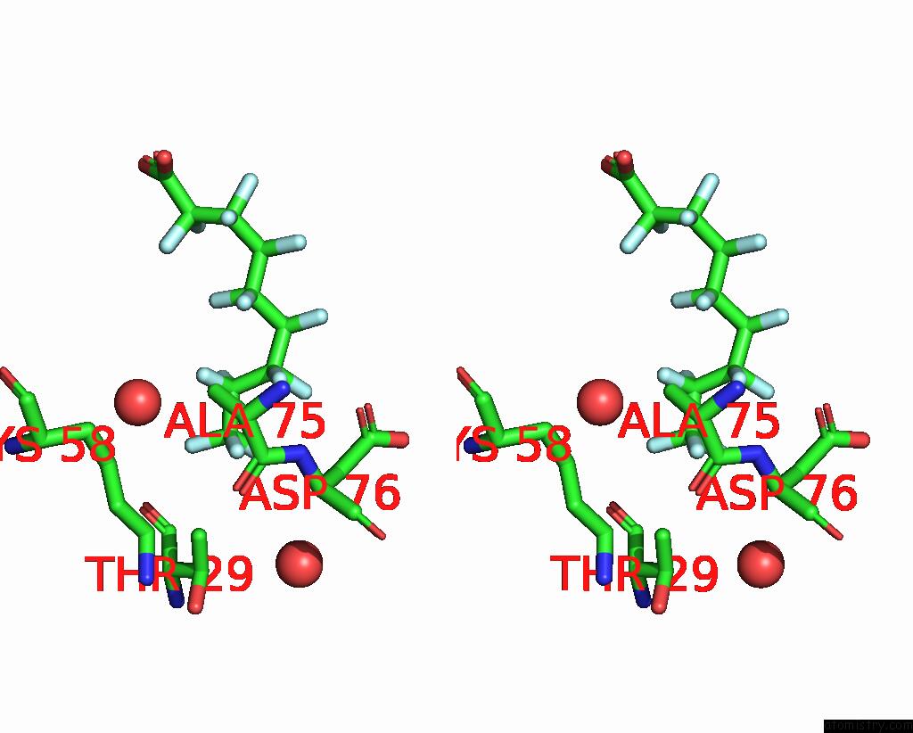
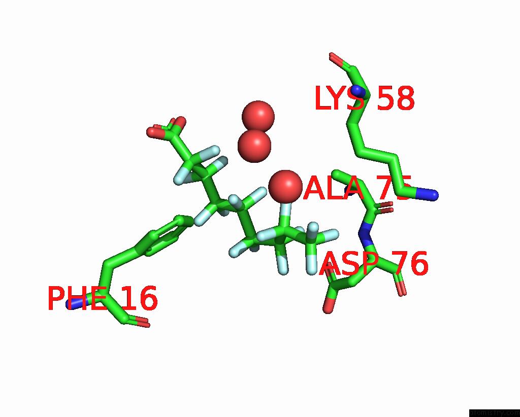
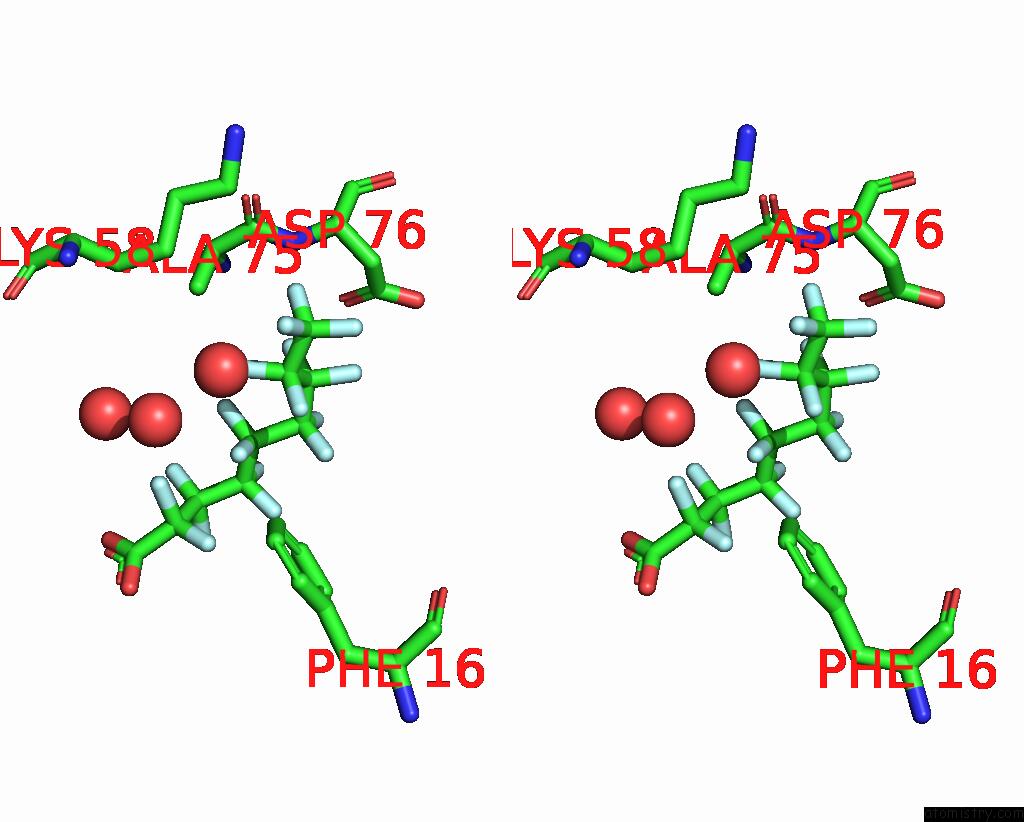
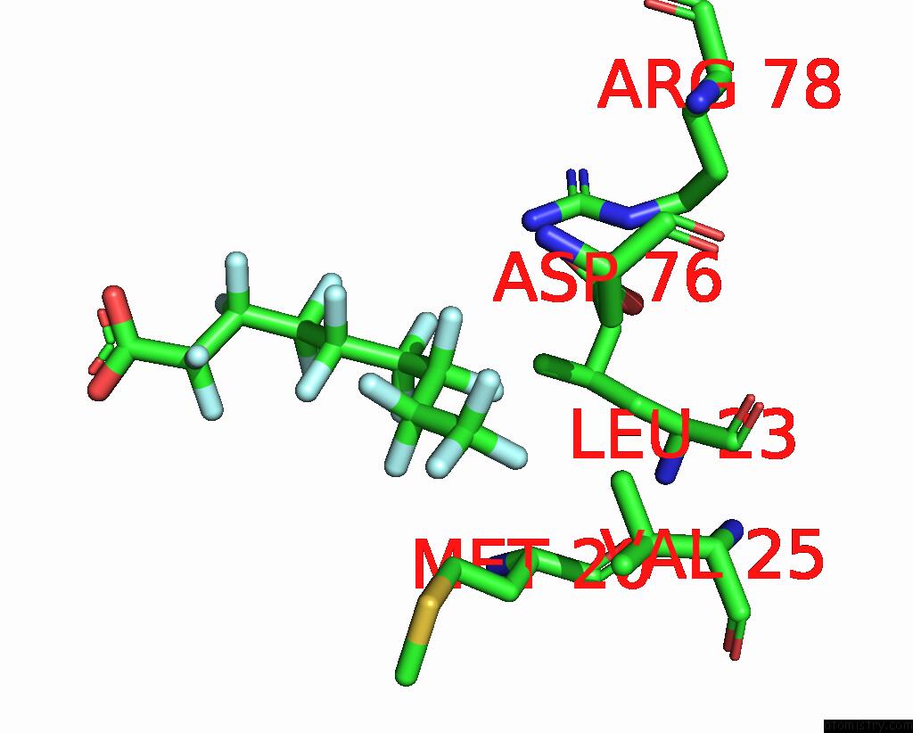
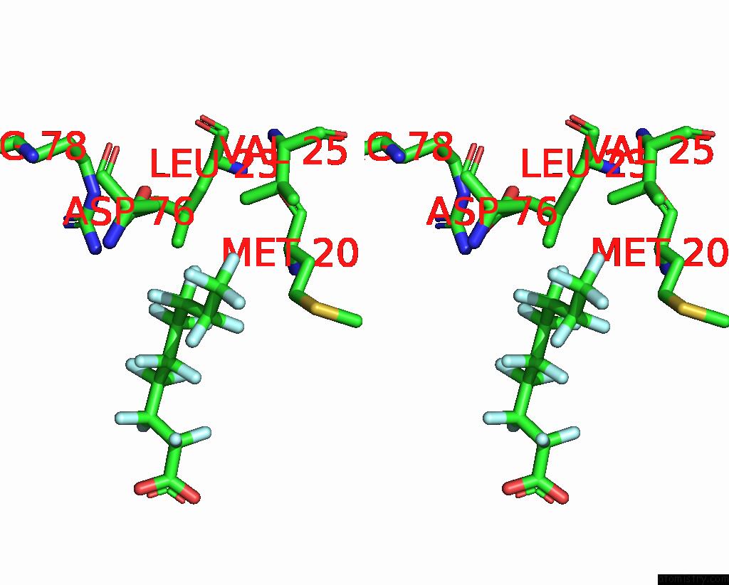
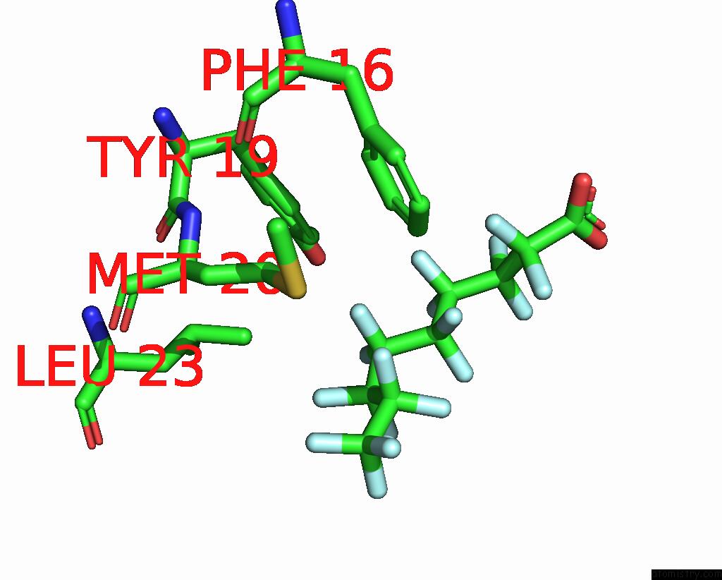
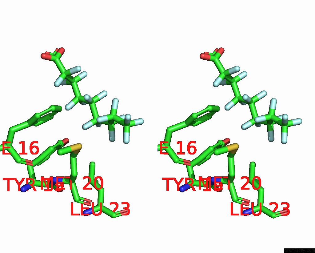
Fluorine binding site 1 out of 17 in 7feu
Go back to
Fluorine binding site 1 out
of 17 in the The 0.95 Angstrom X-Ray Structure of the Human Heart Fatty Acid- Binding Protein Complexed with Perfluorononanoic Acid
Mono view
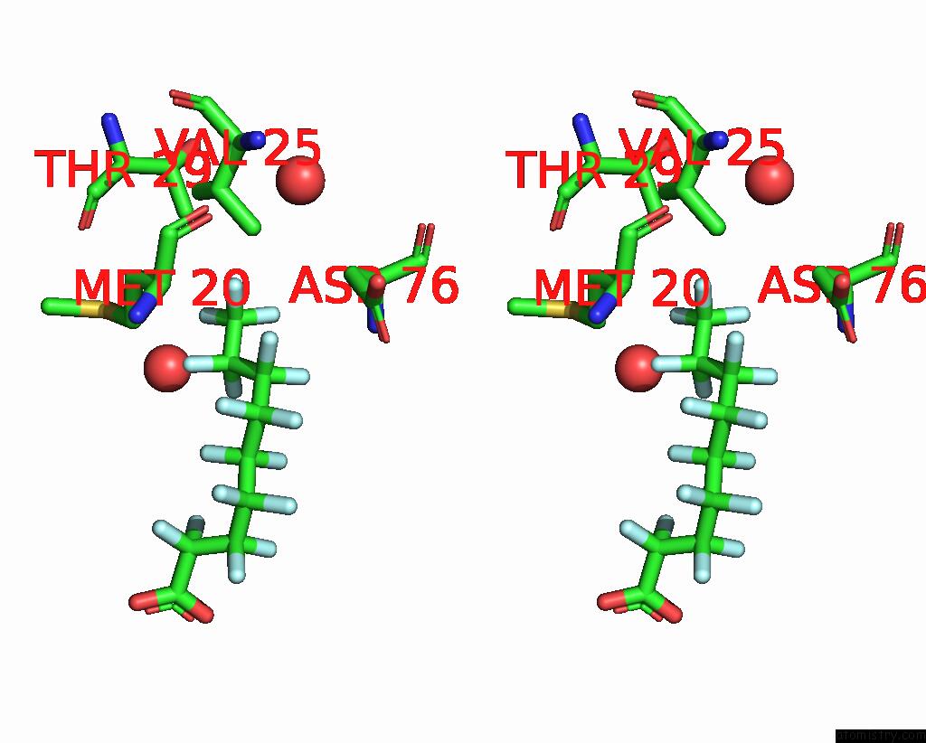
Stereo pair view
Mono view
Stereo pair view
A full contact list of Fluorine with other atoms in the F binding
site number 1 of The 0.95 Angstrom X-Ray Structure of the Human Heart Fatty Acid- Binding Protein Complexed with Perfluorononanoic Acid within 5.0Å range:
|
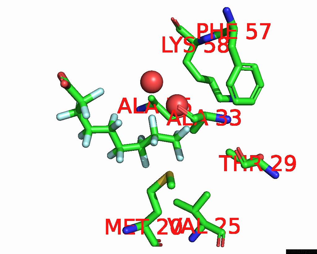
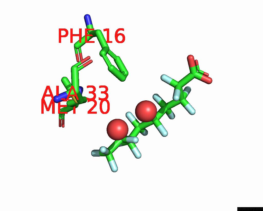
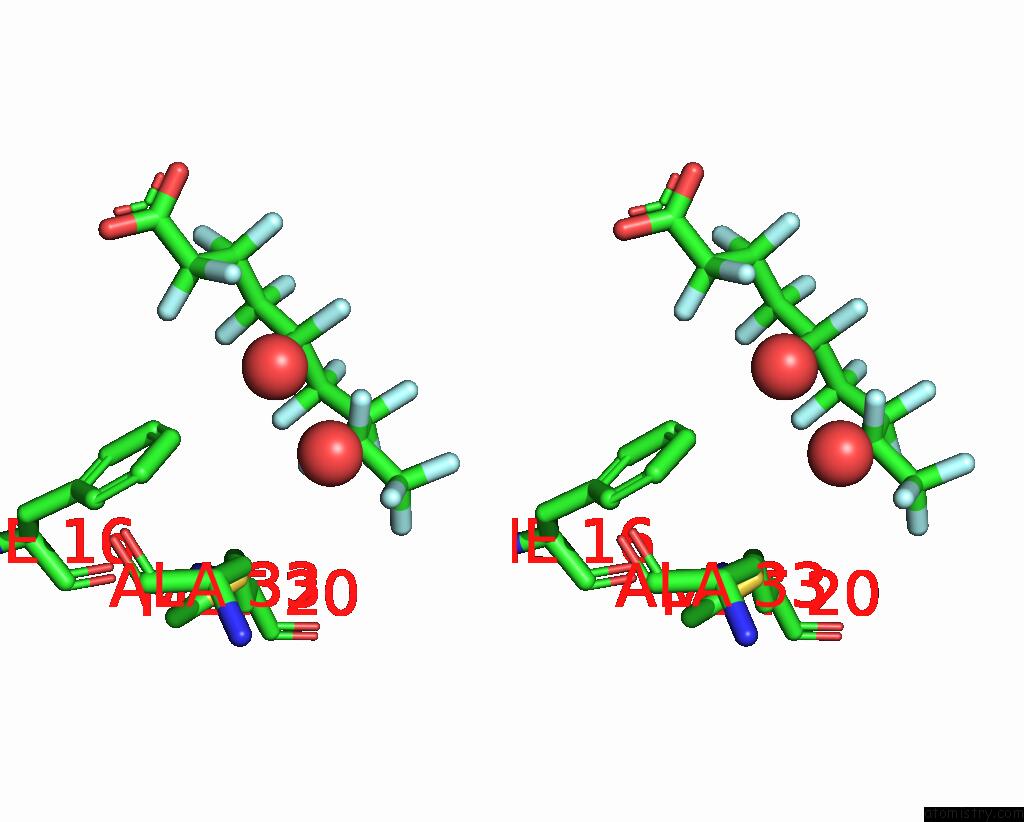
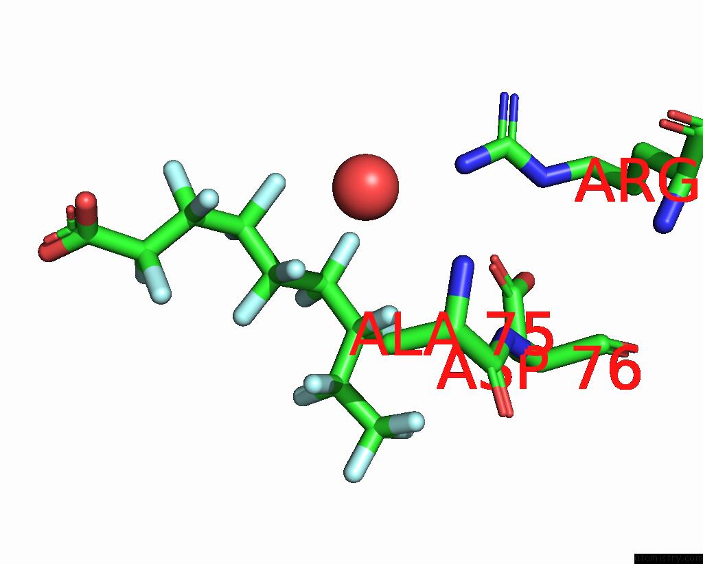
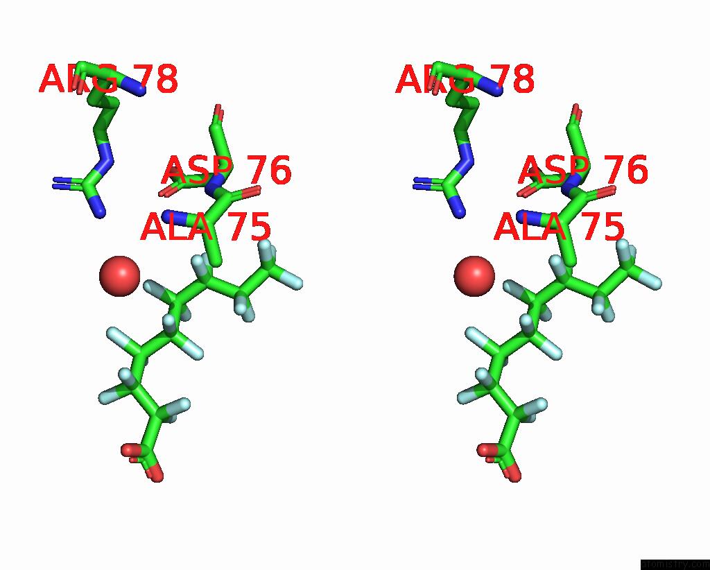
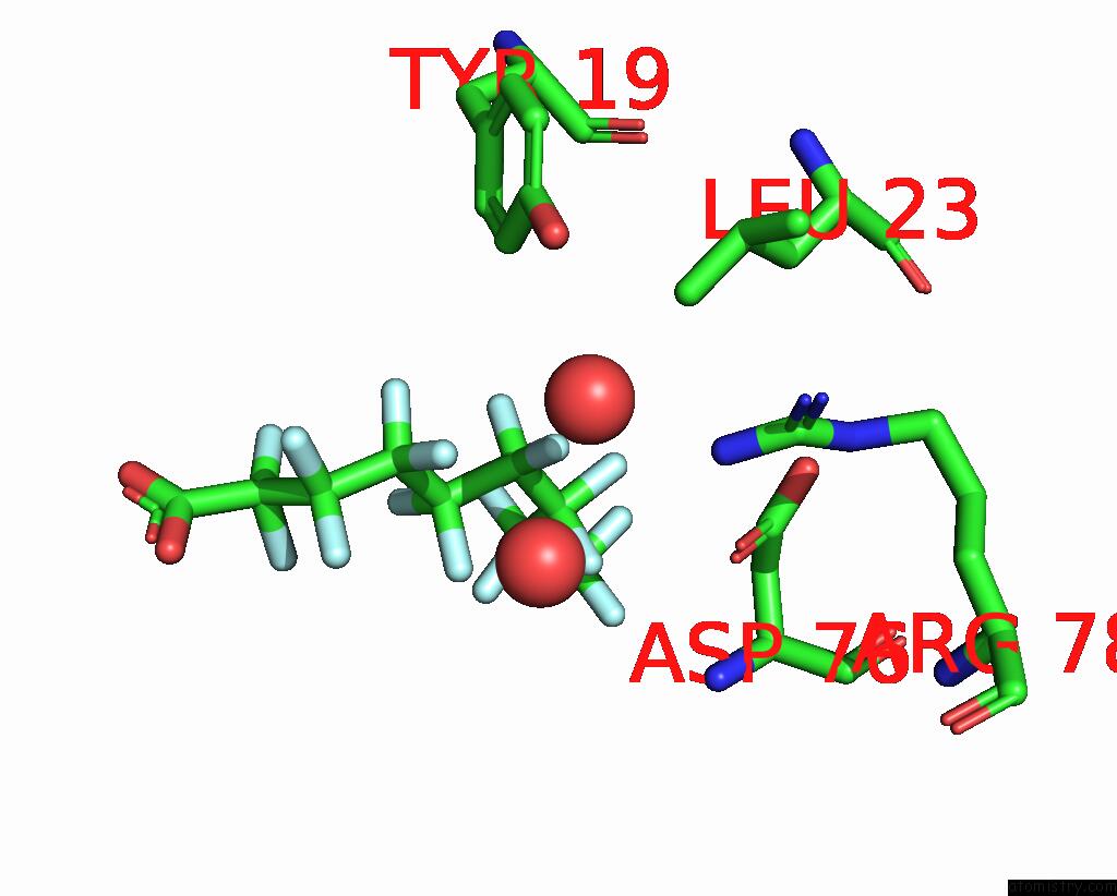
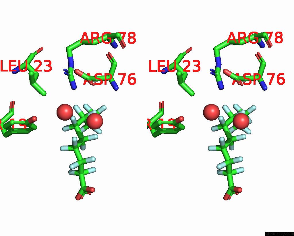
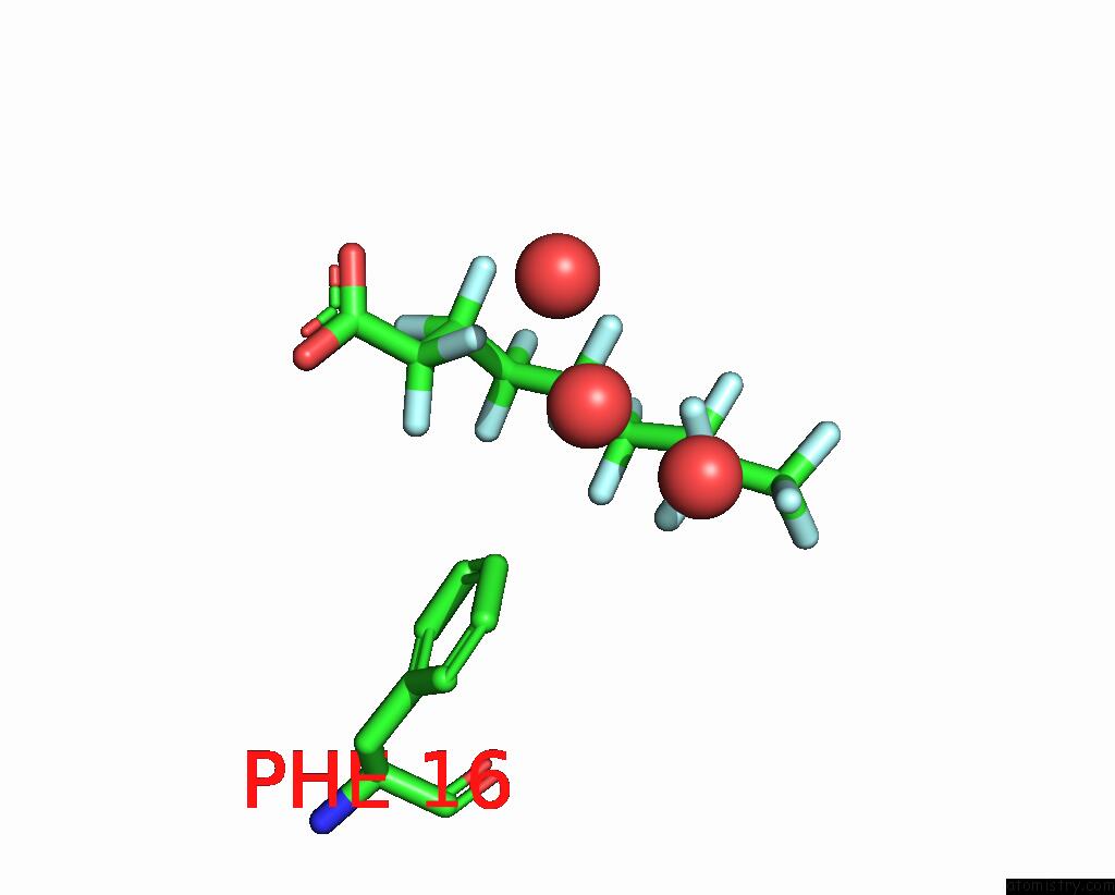
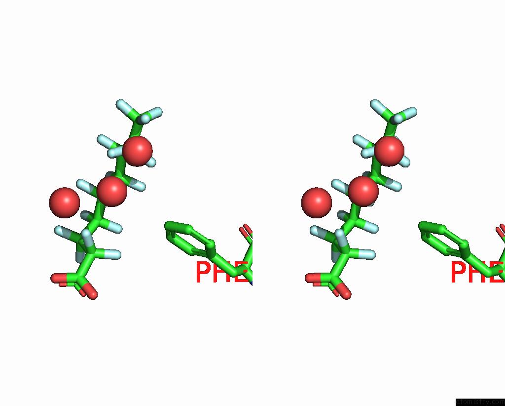
Fluorine binding site 2 out of 17 in 7feu
Go back to
Fluorine binding site 2 out
of 17 in the The 0.95 Angstrom X-Ray Structure of the Human Heart Fatty Acid- Binding Protein Complexed with Perfluorononanoic Acid
Mono view
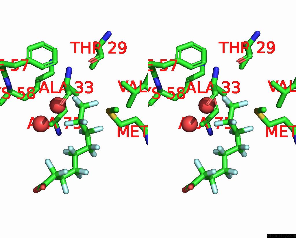
Stereo pair view
Mono view
Stereo pair view
A full contact list of Fluorine with other atoms in the F binding
site number 2 of The 0.95 Angstrom X-Ray Structure of the Human Heart Fatty Acid- Binding Protein Complexed with Perfluorononanoic Acid within 5.0Å range:
|
Fluorine binding site 3 out of 17 in 7feu
Go back to
Fluorine binding site 3 out
of 17 in the The 0.95 Angstrom X-Ray Structure of the Human Heart Fatty Acid- Binding Protein Complexed with Perfluorononanoic Acid
Mono view
Stereo pair view
Mono view
Stereo pair view
A full contact list of Fluorine with other atoms in the F binding
site number 3 of The 0.95 Angstrom X-Ray Structure of the Human Heart Fatty Acid- Binding Protein Complexed with Perfluorononanoic Acid within 5.0Å range:
|
Fluorine binding site 4 out of 17 in 7feu
Go back to
Fluorine binding site 4 out
of 17 in the The 0.95 Angstrom X-Ray Structure of the Human Heart Fatty Acid- Binding Protein Complexed with Perfluorononanoic Acid
Mono view
Stereo pair view
Mono view
Stereo pair view
A full contact list of Fluorine with other atoms in the F binding
site number 4 of The 0.95 Angstrom X-Ray Structure of the Human Heart Fatty Acid- Binding Protein Complexed with Perfluorononanoic Acid within 5.0Å range:
|
Fluorine binding site 5 out of 17 in 7feu
Go back to
Fluorine binding site 5 out
of 17 in the The 0.95 Angstrom X-Ray Structure of the Human Heart Fatty Acid- Binding Protein Complexed with Perfluorononanoic Acid
Mono view
Stereo pair view
Mono view
Stereo pair view
A full contact list of Fluorine with other atoms in the F binding
site number 5 of The 0.95 Angstrom X-Ray Structure of the Human Heart Fatty Acid- Binding Protein Complexed with Perfluorononanoic Acid within 5.0Å range:
|
Fluorine binding site 6 out of 17 in 7feu
Go back to
Fluorine binding site 6 out
of 17 in the The 0.95 Angstrom X-Ray Structure of the Human Heart Fatty Acid- Binding Protein Complexed with Perfluorononanoic Acid
Mono view
Stereo pair view
Mono view
Stereo pair view
A full contact list of Fluorine with other atoms in the F binding
site number 6 of The 0.95 Angstrom X-Ray Structure of the Human Heart Fatty Acid- Binding Protein Complexed with Perfluorononanoic Acid within 5.0Å range:
|
Fluorine binding site 7 out of 17 in 7feu
Go back to
Fluorine binding site 7 out
of 17 in the The 0.95 Angstrom X-Ray Structure of the Human Heart Fatty Acid- Binding Protein Complexed with Perfluorononanoic Acid
Mono view
Stereo pair view
Mono view
Stereo pair view
A full contact list of Fluorine with other atoms in the F binding
site number 7 of The 0.95 Angstrom X-Ray Structure of the Human Heart Fatty Acid- Binding Protein Complexed with Perfluorononanoic Acid within 5.0Å range:
|
Fluorine binding site 8 out of 17 in 7feu
Go back to
Fluorine binding site 8 out
of 17 in the The 0.95 Angstrom X-Ray Structure of the Human Heart Fatty Acid- Binding Protein Complexed with Perfluorononanoic Acid
Mono view
Stereo pair view
Mono view
Stereo pair view
A full contact list of Fluorine with other atoms in the F binding
site number 8 of The 0.95 Angstrom X-Ray Structure of the Human Heart Fatty Acid- Binding Protein Complexed with Perfluorononanoic Acid within 5.0Å range:
|
Fluorine binding site 9 out of 17 in 7feu
Go back to
Fluorine binding site 9 out
of 17 in the The 0.95 Angstrom X-Ray Structure of the Human Heart Fatty Acid- Binding Protein Complexed with Perfluorononanoic Acid
Mono view
Stereo pair view
Mono view
Stereo pair view
A full contact list of Fluorine with other atoms in the F binding
site number 9 of The 0.95 Angstrom X-Ray Structure of the Human Heart Fatty Acid- Binding Protein Complexed with Perfluorononanoic Acid within 5.0Å range:
|
Fluorine binding site 10 out of 17 in 7feu
Go back to
Fluorine binding site 10 out
of 17 in the The 0.95 Angstrom X-Ray Structure of the Human Heart Fatty Acid- Binding Protein Complexed with Perfluorononanoic Acid
Mono view
Stereo pair view
Mono view
Stereo pair view
A full contact list of Fluorine with other atoms in the F binding
site number 10 of The 0.95 Angstrom X-Ray Structure of the Human Heart Fatty Acid- Binding Protein Complexed with Perfluorononanoic Acid within 5.0Å range:
|
Reference:
S.Sugiyama,
K.Kakinouchi,
T.Hara,
S.Matsuoka,
H.Tsuchikawa,
M.Sonoyama,
Y.Inoue,
F.Hayashi,
M.Murata.
The 0.95 Angstrom X-Ray Structure of the Human Heart Fatty Acid-Binding Protein Complexed with Perfluorononanoic Acid To Be Published.
Page generated: Tue Jul 15 19:27:30 2025
Last articles
Fe in 9UD4Fe in 9UD5
Fe in 9UD3
Fe in 9QM1
Fe in 9UD2
Fe in 9U5G
Fe in 9QM0
Fe in 9R7J
Fe in 9NF0
Fe in 9NEZ