Fluorine »
PDB 7kqd-7kzy »
7kvu »
Fluorine in PDB 7kvu: Crystal Structure of Squash Rna Aptamer in Complex with Dfhbi-1T
Protein crystallography data
The structure of Crystal Structure of Squash Rna Aptamer in Complex with Dfhbi-1T, PDB code: 7kvu
was solved by
L.Truong,
A.R.Ferre-D'amare,
with X-Ray Crystallography technique. A brief refinement statistics is given in the table below:
| Resolution Low / High (Å) | 46.46 / 2.68 |
| Space group | P 42 21 2 |
| Cell size a, b, c (Å), α, β, γ (°) | 103.731, 103.731, 51.959, 90, 90, 90 |
| R / Rfree (%) | 20.9 / 24.7 |
Other elements in 7kvu:
The structure of Crystal Structure of Squash Rna Aptamer in Complex with Dfhbi-1T also contains other interesting chemical elements:
| Potassium | (K) | 3 atoms |
| Magnesium | (Mg) | 4 atoms |
Fluorine Binding Sites:
The binding sites of Fluorine atom in the Crystal Structure of Squash Rna Aptamer in Complex with Dfhbi-1T
(pdb code 7kvu). This binding sites where shown within
5.0 Angstroms radius around Fluorine atom.
In total 5 binding sites of Fluorine where determined in the Crystal Structure of Squash Rna Aptamer in Complex with Dfhbi-1T, PDB code: 7kvu:
Jump to Fluorine binding site number: 1; 2; 3; 4; 5;
In total 5 binding sites of Fluorine where determined in the Crystal Structure of Squash Rna Aptamer in Complex with Dfhbi-1T, PDB code: 7kvu:
Jump to Fluorine binding site number: 1; 2; 3; 4; 5;
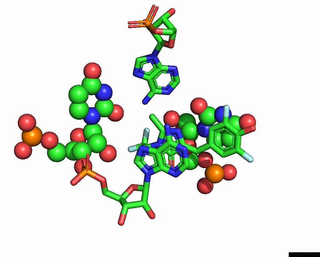
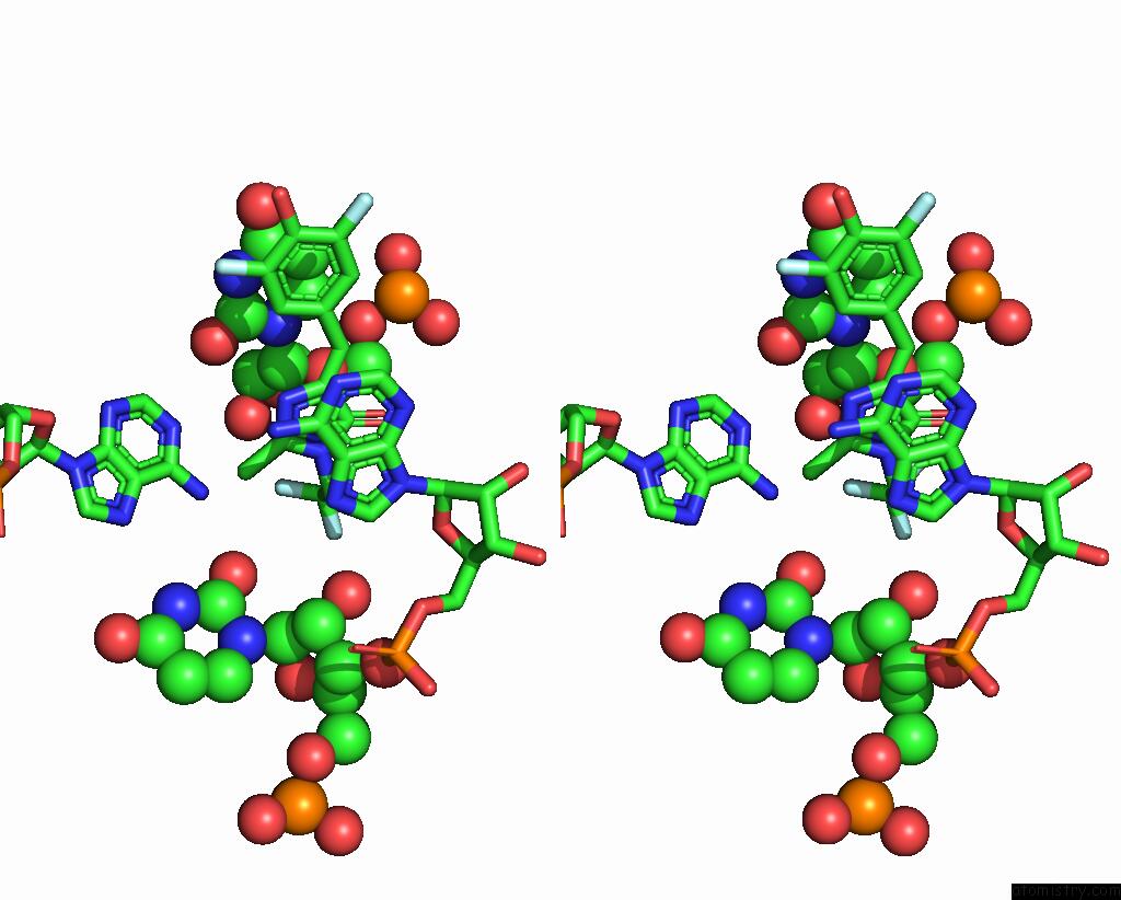
Fluorine binding site 1 out of 5 in 7kvu
Go back to
Fluorine binding site 1 out
of 5 in the Crystal Structure of Squash Rna Aptamer in Complex with Dfhbi-1T
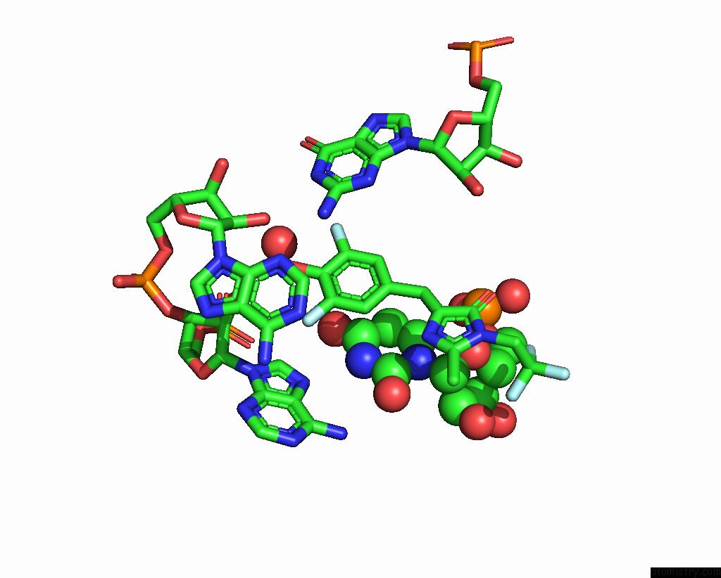
Mono view
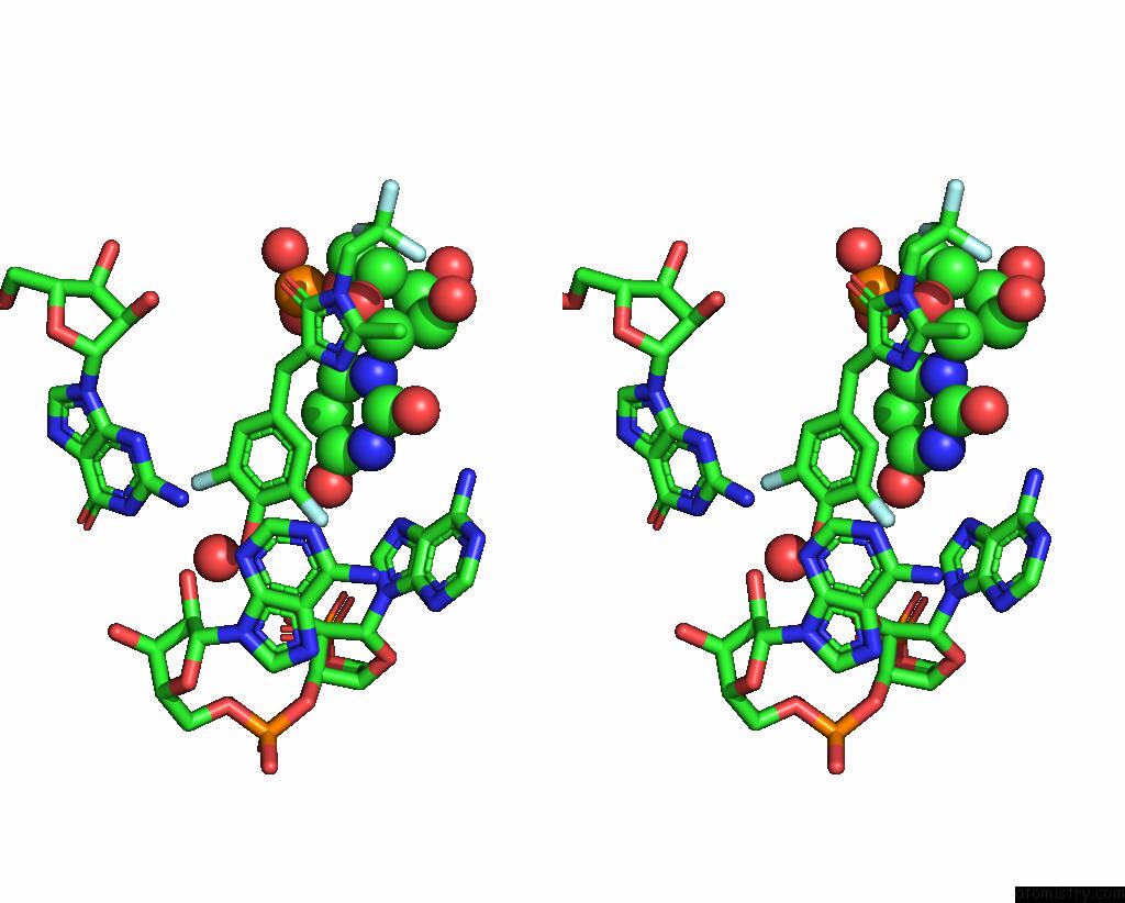
Stereo pair view
Mono view
Stereo pair view
A full contact list of Fluorine with other atoms in the F binding
site number 1 of Crystal Structure of Squash Rna Aptamer in Complex with Dfhbi-1T within 5.0Å range:
|
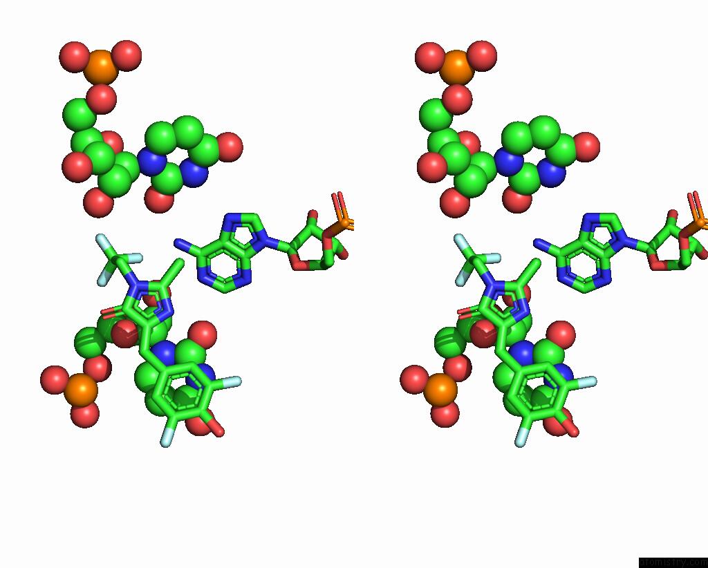
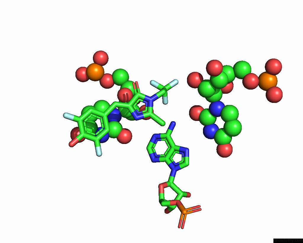
Fluorine binding site 2 out of 5 in 7kvu
Go back to
Fluorine binding site 2 out
of 5 in the Crystal Structure of Squash Rna Aptamer in Complex with Dfhbi-1T
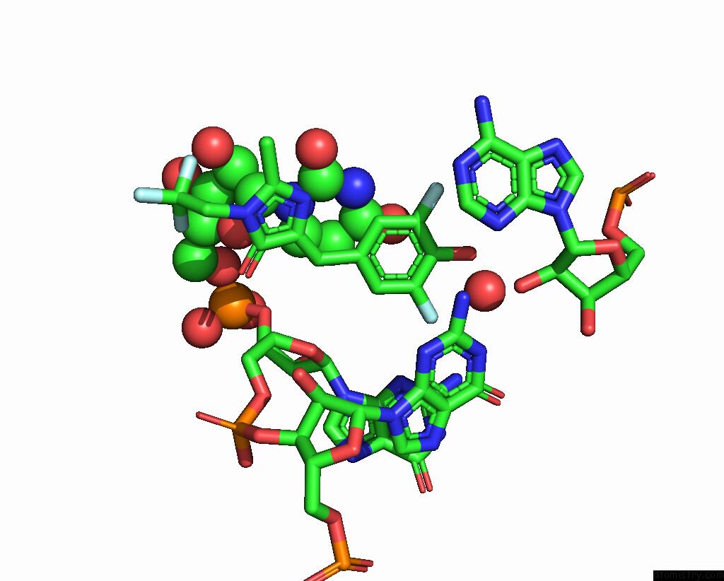
Mono view

Stereo pair view
Mono view
Stereo pair view
A full contact list of Fluorine with other atoms in the F binding
site number 2 of Crystal Structure of Squash Rna Aptamer in Complex with Dfhbi-1T within 5.0Å range:
|
Fluorine binding site 3 out of 5 in 7kvu
Go back to
Fluorine binding site 3 out
of 5 in the Crystal Structure of Squash Rna Aptamer in Complex with Dfhbi-1T

Mono view
Stereo pair view
Mono view
Stereo pair view
A full contact list of Fluorine with other atoms in the F binding
site number 3 of Crystal Structure of Squash Rna Aptamer in Complex with Dfhbi-1T within 5.0Å range:
|
Fluorine binding site 4 out of 5 in 7kvu
Go back to
Fluorine binding site 4 out
of 5 in the Crystal Structure of Squash Rna Aptamer in Complex with Dfhbi-1T
Mono view
Stereo pair view
Mono view
Stereo pair view
A full contact list of Fluorine with other atoms in the F binding
site number 4 of Crystal Structure of Squash Rna Aptamer in Complex with Dfhbi-1T within 5.0Å range:
|
Fluorine binding site 5 out of 5 in 7kvu
Go back to
Fluorine binding site 5 out
of 5 in the Crystal Structure of Squash Rna Aptamer in Complex with Dfhbi-1T
Mono view

Stereo pair view
Mono view
Stereo pair view
A full contact list of Fluorine with other atoms in the F binding
site number 5 of Crystal Structure of Squash Rna Aptamer in Complex with Dfhbi-1T within 5.0Å range:
|
Reference:
L.Truong,
H.Kooshapur,
S.K.Dey,
X.Li,
N.Tjandra,
S.R.Jaffrey,
A.R.Ferre-D'amare.
The Fluorescent Aptamer Squash Extensively Repurposes the Adenine Riboswitch Fold. Nat.Chem.Biol. V. 18 191 2022.
ISSN: ESSN 1552-4469
PubMed: 34937911
DOI: 10.1038/S41589-021-00931-2
Page generated: Tue Jul 15 20:57:15 2025
ISSN: ESSN 1552-4469
PubMed: 34937911
DOI: 10.1038/S41589-021-00931-2
Last articles
K in 8GABK in 8G7M
K in 8G6X
K in 8G2N
K in 8G20
K in 8FS1
K in 8FS2
K in 8G1Z
K in 8FZ7
K in 8FZU