Fluorine »
PDB 7qmw-7r9f »
7qu0 »
Fluorine in PDB 7qu0: X-Ray Structure of Fad Domain of Nqrf of Klebsiella Pneumoniae
Enzymatic activity of X-Ray Structure of Fad Domain of Nqrf of Klebsiella Pneumoniae
All present enzymatic activity of X-Ray Structure of Fad Domain of Nqrf of Klebsiella Pneumoniae:
7.2.1.1;
7.2.1.1;
Protein crystallography data
The structure of X-Ray Structure of Fad Domain of Nqrf of Klebsiella Pneumoniae, PDB code: 7qu0
was solved by
D.Stegmann,
J.Steuber,
G.Fritz,
with X-Ray Crystallography technique. A brief refinement statistics is given in the table below:
| Resolution Low / High (Å) | 43.18 / 1.62 |
| Space group | P 41 21 2 |
| Cell size a, b, c (Å), α, β, γ (°) | 98.84, 98.84, 88.75, 90, 90, 90 |
| R / Rfree (%) | 16.8 / 19.3 |
Fluorine Binding Sites:
The binding sites of Fluorine atom in the X-Ray Structure of Fad Domain of Nqrf of Klebsiella Pneumoniae
(pdb code 7qu0). This binding sites where shown within
5.0 Angstroms radius around Fluorine atom.
In total 2 binding sites of Fluorine where determined in the X-Ray Structure of Fad Domain of Nqrf of Klebsiella Pneumoniae, PDB code: 7qu0:
Jump to Fluorine binding site number: 1; 2;
In total 2 binding sites of Fluorine where determined in the X-Ray Structure of Fad Domain of Nqrf of Klebsiella Pneumoniae, PDB code: 7qu0:
Jump to Fluorine binding site number: 1; 2;
Fluorine binding site 1 out of 2 in 7qu0
Go back to
Fluorine binding site 1 out
of 2 in the X-Ray Structure of Fad Domain of Nqrf of Klebsiella Pneumoniae
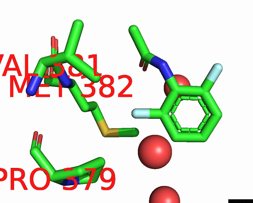
Mono view
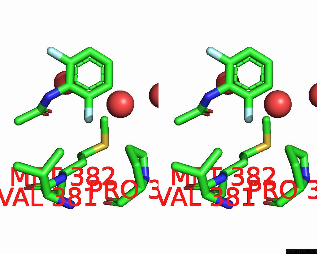
Stereo pair view
Mono view
Stereo pair view
A full contact list of Fluorine with other atoms in the F binding
site number 1 of X-Ray Structure of Fad Domain of Nqrf of Klebsiella Pneumoniae within 5.0Å range:
|
Fluorine binding site 2 out of 2 in 7qu0
Go back to
Fluorine binding site 2 out
of 2 in the X-Ray Structure of Fad Domain of Nqrf of Klebsiella Pneumoniae

Mono view
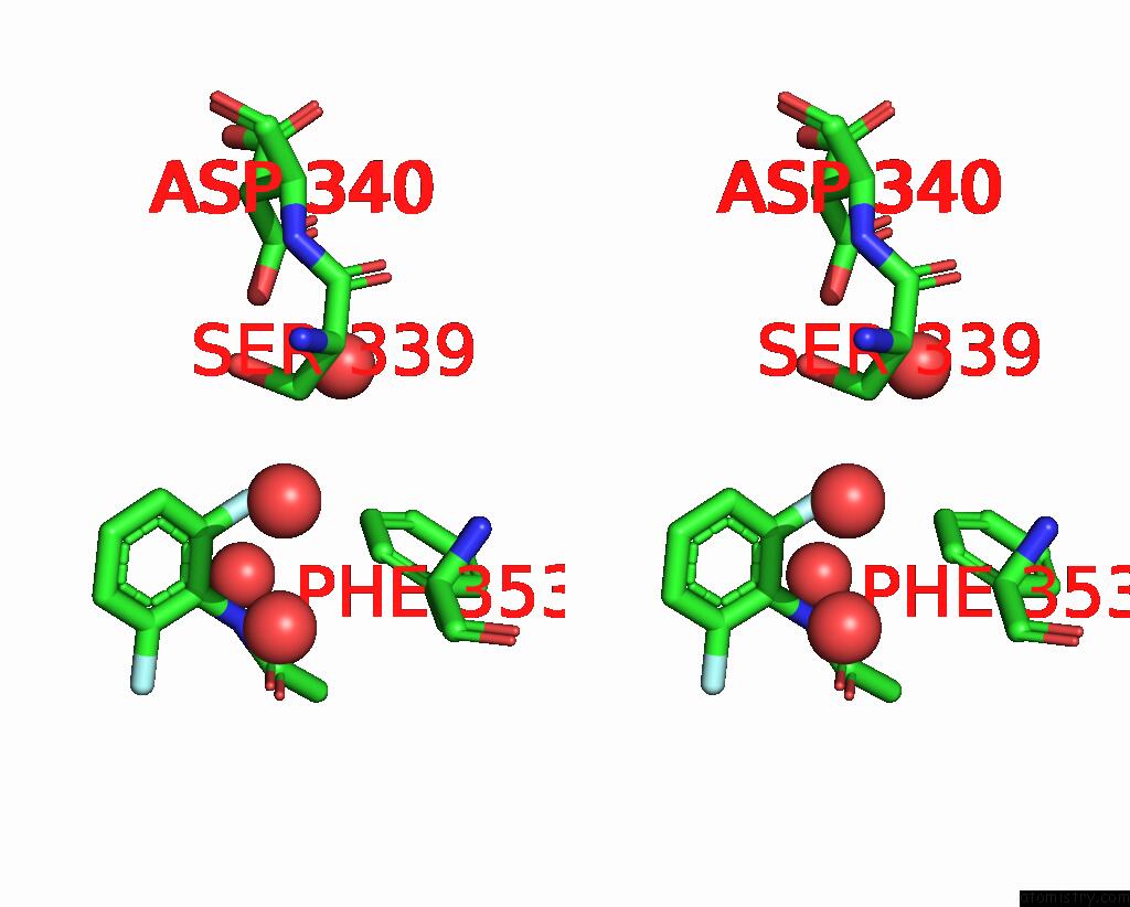
Stereo pair view
Mono view
Stereo pair view
A full contact list of Fluorine with other atoms in the F binding
site number 2 of X-Ray Structure of Fad Domain of Nqrf of Klebsiella Pneumoniae within 5.0Å range:
|
Reference:
J.W.Kaminski,
L.Vera,
D.P.Stegmann,
J.Vering,
D.Eris,
K.M.L.Smith,
C.Y.Huang,
N.Meier,
J.Steuber,
M.Wang,
G.Fritz,
J.A.Wojdyla,
M.E.Sharpe.
Fast Fragment- and Compound-Screening Pipeline at the Swiss Light Source. Acta Crystallogr D Struct V. 78 328 2022BIOL.
ISSN: ISSN 2059-7983
PubMed: 35234147
DOI: 10.1107/S2059798322000705
Page generated: Tue Jul 15 23:19:17 2025
ISSN: ISSN 2059-7983
PubMed: 35234147
DOI: 10.1107/S2059798322000705
Last articles
Na in 4WCXNa in 4WET
Na in 4WCN
Na in 4WED
Na in 4W5Z
Na in 4W9N
Na in 4WCL
Na in 4W96
Na in 4W94
Na in 4W78