Fluorine »
PDB 8b99-8by9 »
8bm2 »
Fluorine in PDB 8bm2: Crystal Structure of JAK2 JH1 in Complex with Gandotinib
Enzymatic activity of Crystal Structure of JAK2 JH1 in Complex with Gandotinib
All present enzymatic activity of Crystal Structure of JAK2 JH1 in Complex with Gandotinib:
2.7.10.2;
2.7.10.2;
Protein crystallography data
The structure of Crystal Structure of JAK2 JH1 in Complex with Gandotinib, PDB code: 8bm2
was solved by
Y.Miao,
T.Haikarainen,
with X-Ray Crystallography technique. A brief refinement statistics is given in the table below:
| Resolution Low / High (Å) | 58.46 / 1.50 |
| Space group | I 1 2 1 |
| Cell size a, b, c (Å), α, β, γ (°) | 99.894, 69.254, 110.299, 90, 98.68, 90 |
| R / Rfree (%) | 17.7 / 20.2 |
Other elements in 8bm2:
The structure of Crystal Structure of JAK2 JH1 in Complex with Gandotinib also contains other interesting chemical elements:
| Chlorine | (Cl) | 2 atoms |
Fluorine Binding Sites:
The binding sites of Fluorine atom in the Crystal Structure of JAK2 JH1 in Complex with Gandotinib
(pdb code 8bm2). This binding sites where shown within
5.0 Angstroms radius around Fluorine atom.
In total 2 binding sites of Fluorine where determined in the Crystal Structure of JAK2 JH1 in Complex with Gandotinib, PDB code: 8bm2:
Jump to Fluorine binding site number: 1; 2;
In total 2 binding sites of Fluorine where determined in the Crystal Structure of JAK2 JH1 in Complex with Gandotinib, PDB code: 8bm2:
Jump to Fluorine binding site number: 1; 2;
Fluorine binding site 1 out of 2 in 8bm2
Go back to
Fluorine binding site 1 out
of 2 in the Crystal Structure of JAK2 JH1 in Complex with Gandotinib
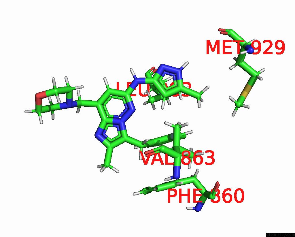
Mono view

Stereo pair view
Mono view
Stereo pair view
A full contact list of Fluorine with other atoms in the F binding
site number 1 of Crystal Structure of JAK2 JH1 in Complex with Gandotinib within 5.0Å range:
|
Fluorine binding site 2 out of 2 in 8bm2
Go back to
Fluorine binding site 2 out
of 2 in the Crystal Structure of JAK2 JH1 in Complex with Gandotinib
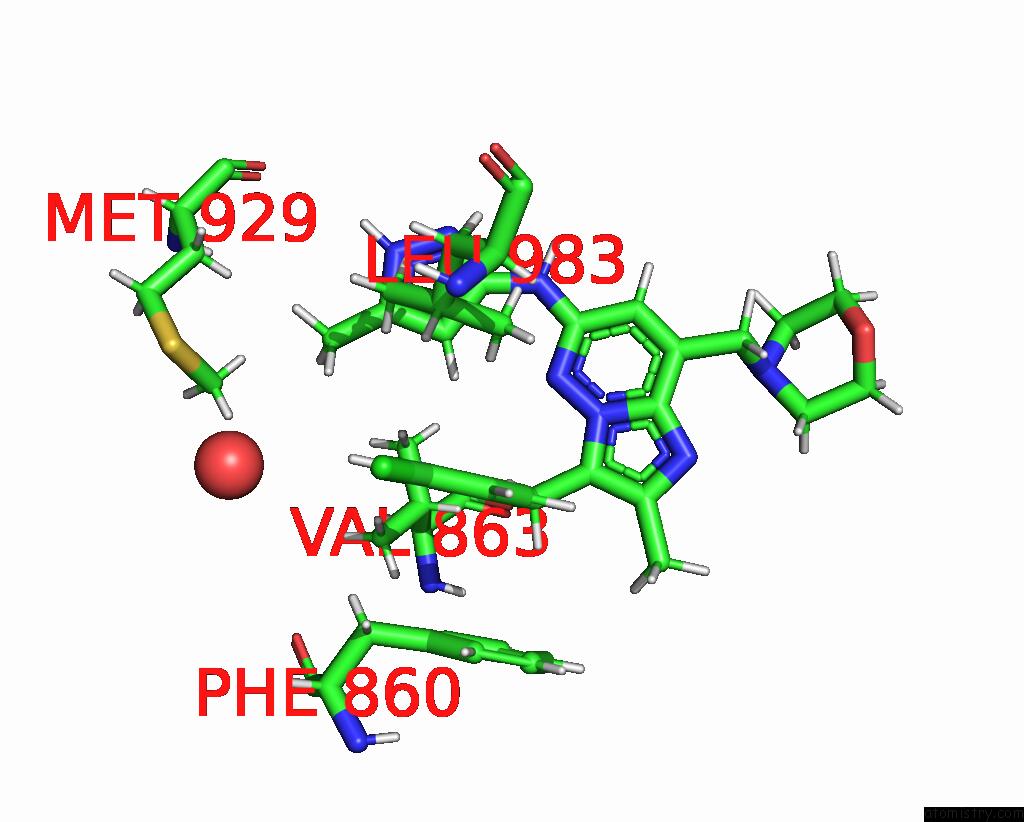
Mono view

Stereo pair view
Mono view
Stereo pair view
A full contact list of Fluorine with other atoms in the F binding
site number 2 of Crystal Structure of JAK2 JH1 in Complex with Gandotinib within 5.0Å range:
|
Reference:
Y.Miao,
O.Silvennoinen,
T.Haikarainen.
Structural Basis For JAK2 Inhibition By Clinical Stage Inhibitors To Be Published.
Page generated: Wed Jul 16 02:55:49 2025
Last articles
Mg in 5H1YMg in 5H1B
Mg in 5H1C
Mg in 5H0V
Mg in 5GZA
Mg in 5GZ9
Mg in 5GYN
Mg in 5GXV
Mg in 5GXT
Mg in 5GX5