Fluorine »
PDB 8dxi-8e90 »
8e25 »
Fluorine in PDB 8e25: Crystal Structure of Sars-Cov-2 Main Protease M49I Mutant in Complex with Nirmatrelvir
Protein crystallography data
The structure of Crystal Structure of Sars-Cov-2 Main Protease M49I Mutant in Complex with Nirmatrelvir, PDB code: 8e25
was solved by
G.D.Noske,
A.S.Godoy,
G.Oliva,
with X-Ray Crystallography technique. A brief refinement statistics is given in the table below:
| Resolution Low / High (Å) | 72.06 / 1.87 |
| Space group | P 21 21 21 |
| Cell size a, b, c (Å), α, β, γ (°) | 67.859, 100.068, 103.533, 90, 90, 90 |
| R / Rfree (%) | 19.4 / 24 |
Fluorine Binding Sites:
The binding sites of Fluorine atom in the Crystal Structure of Sars-Cov-2 Main Protease M49I Mutant in Complex with Nirmatrelvir
(pdb code 8e25). This binding sites where shown within
5.0 Angstroms radius around Fluorine atom.
In total 6 binding sites of Fluorine where determined in the Crystal Structure of Sars-Cov-2 Main Protease M49I Mutant in Complex with Nirmatrelvir, PDB code: 8e25:
Jump to Fluorine binding site number: 1; 2; 3; 4; 5; 6;
In total 6 binding sites of Fluorine where determined in the Crystal Structure of Sars-Cov-2 Main Protease M49I Mutant in Complex with Nirmatrelvir, PDB code: 8e25:
Jump to Fluorine binding site number: 1; 2; 3; 4; 5; 6;
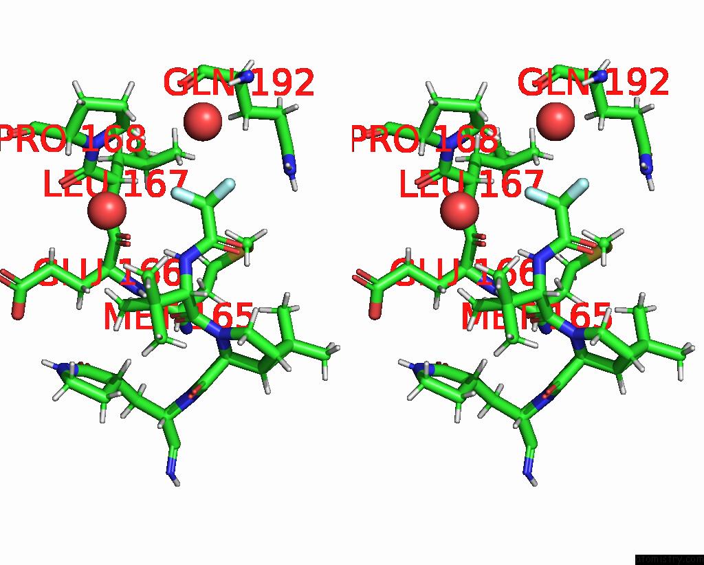
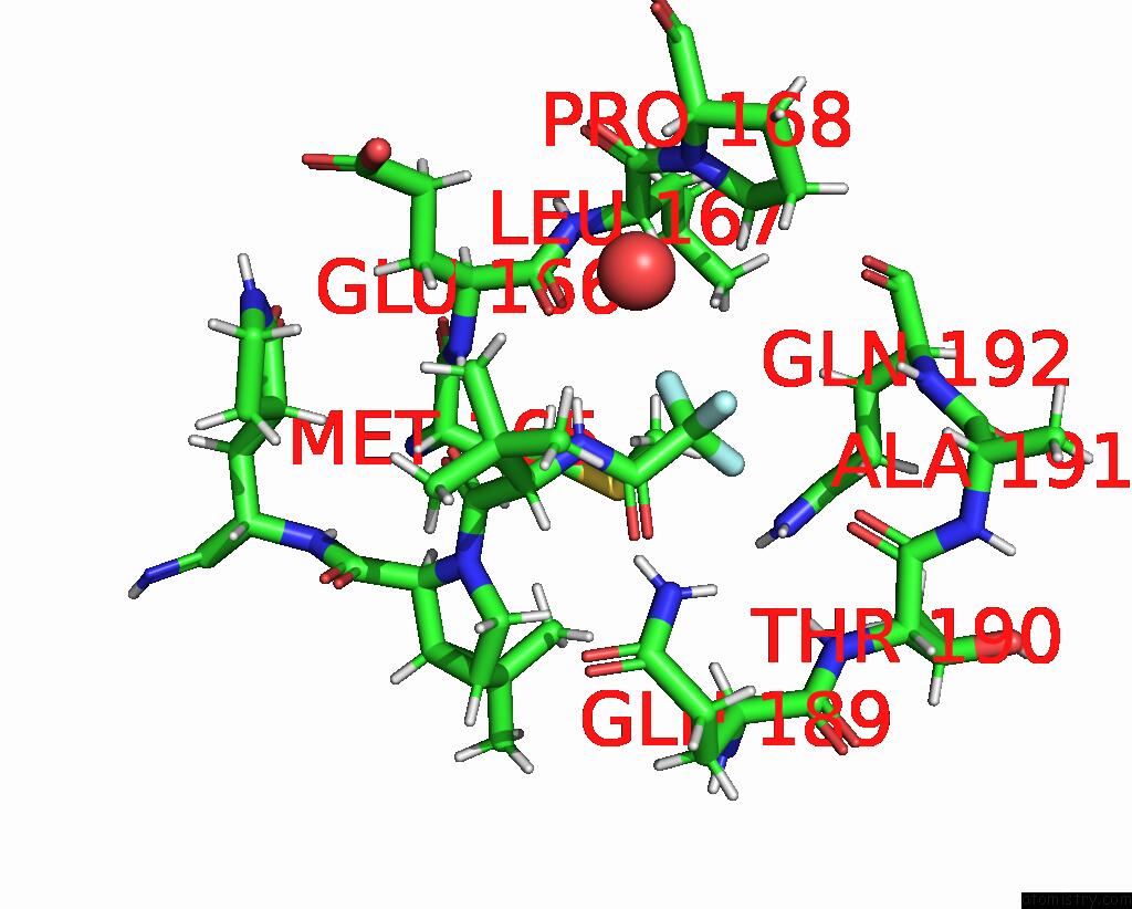
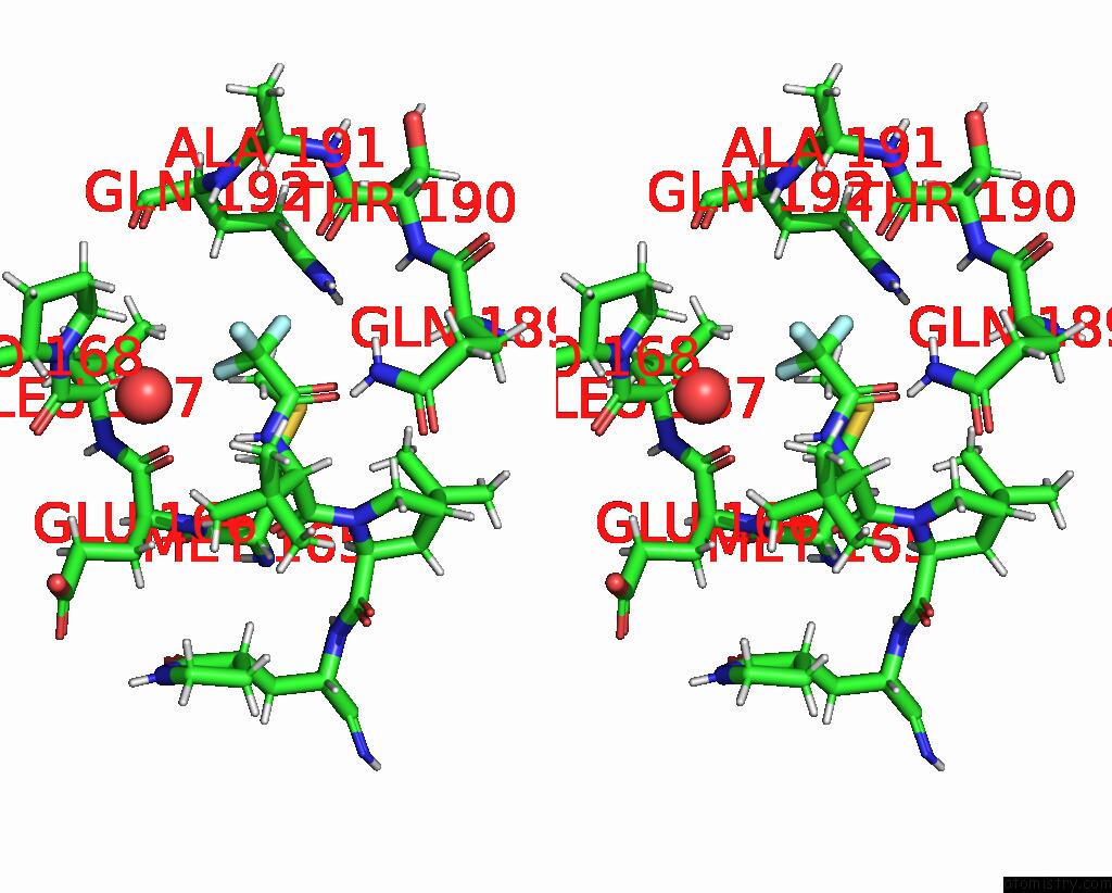
Fluorine binding site 1 out of 6 in 8e25
Go back to
Fluorine binding site 1 out
of 6 in the Crystal Structure of Sars-Cov-2 Main Protease M49I Mutant in Complex with Nirmatrelvir
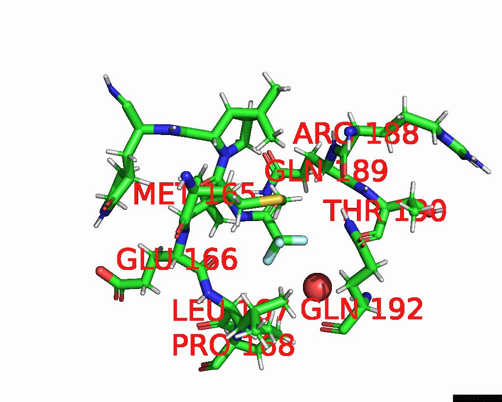
Mono view

Stereo pair view
Mono view
Stereo pair view
A full contact list of Fluorine with other atoms in the F binding
site number 1 of Crystal Structure of Sars-Cov-2 Main Protease M49I Mutant in Complex with Nirmatrelvir within 5.0Å range:
|
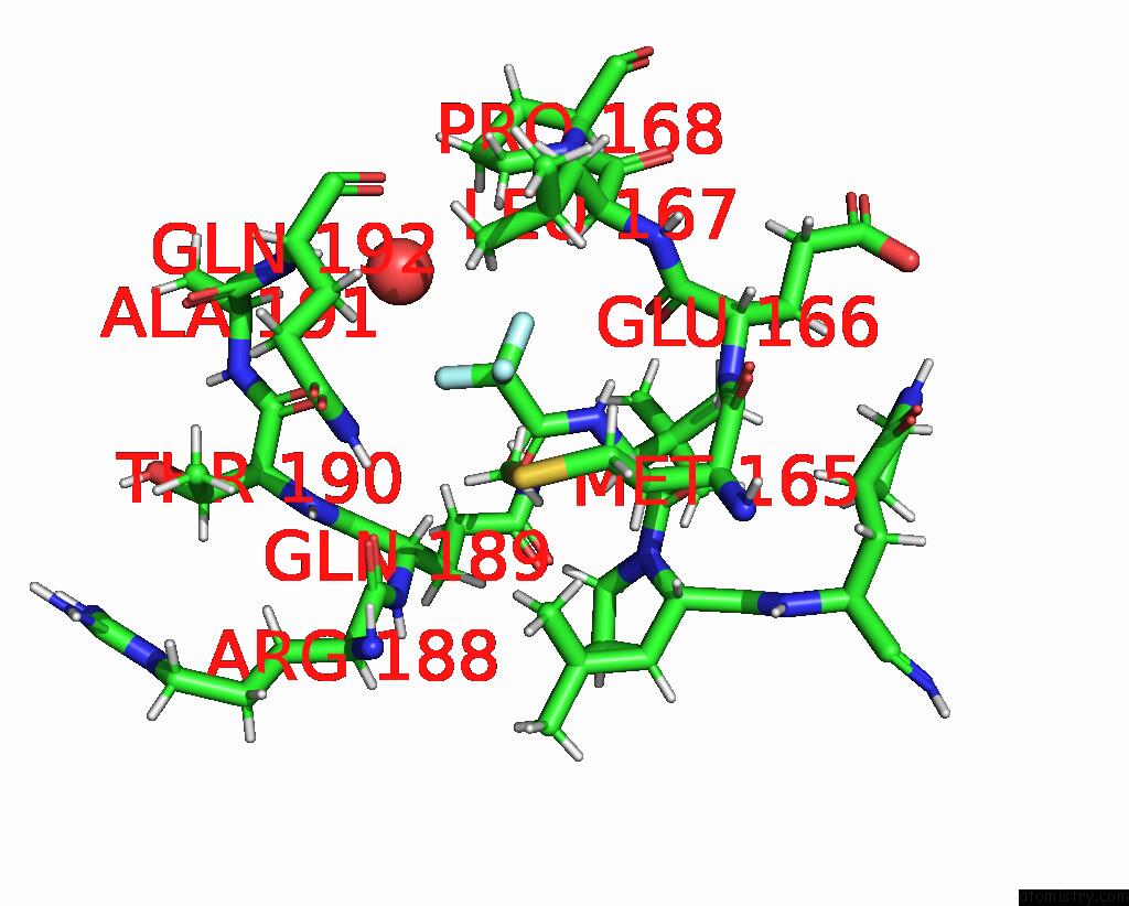
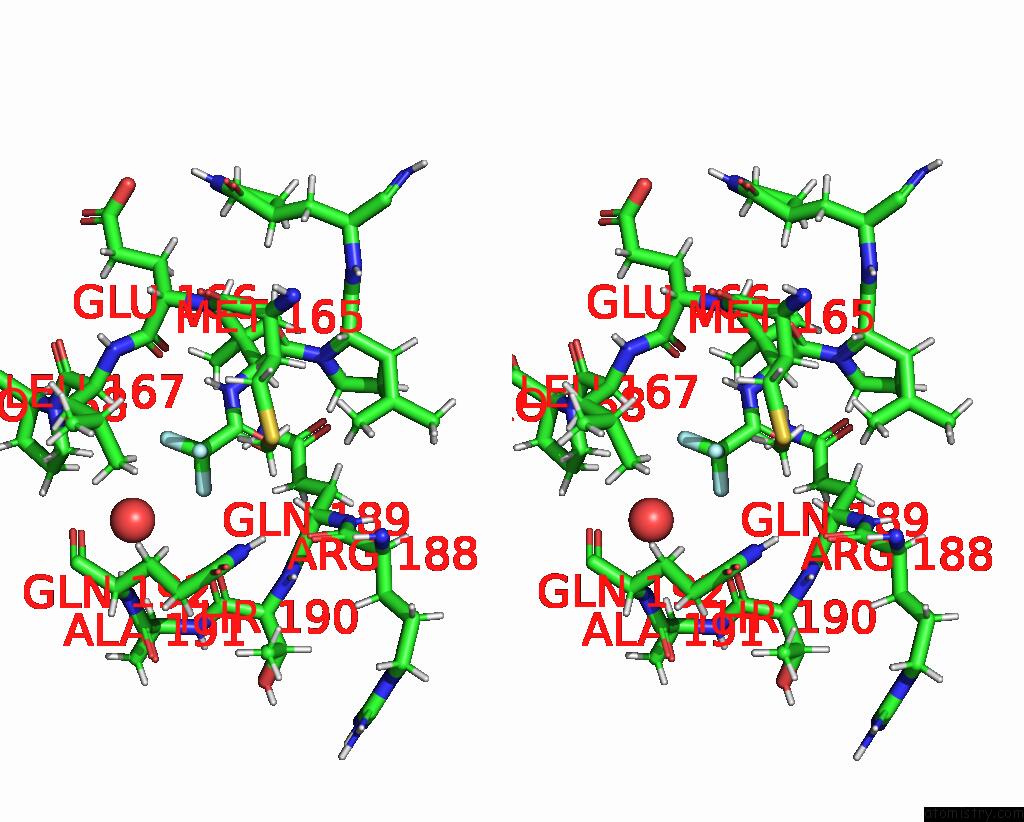
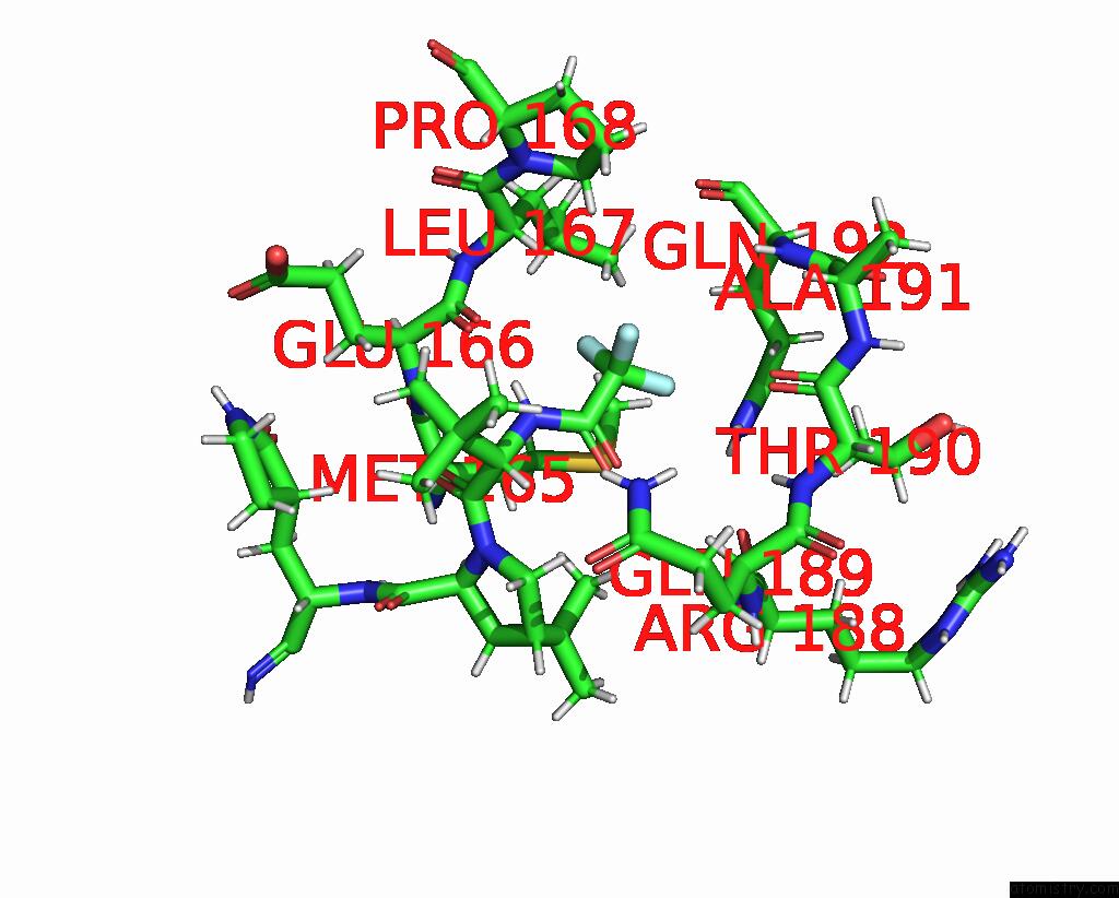
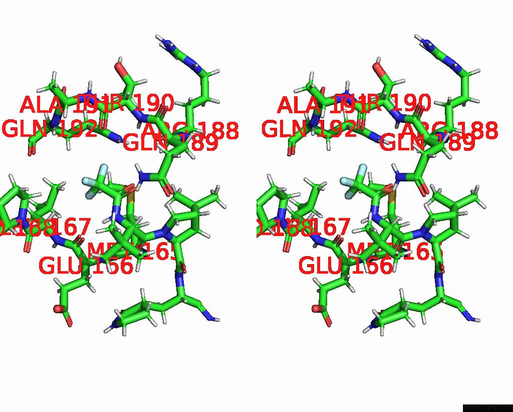
Fluorine binding site 2 out of 6 in 8e25
Go back to
Fluorine binding site 2 out
of 6 in the Crystal Structure of Sars-Cov-2 Main Protease M49I Mutant in Complex with Nirmatrelvir

Mono view
Stereo pair view
Mono view
Stereo pair view
A full contact list of Fluorine with other atoms in the F binding
site number 2 of Crystal Structure of Sars-Cov-2 Main Protease M49I Mutant in Complex with Nirmatrelvir within 5.0Å range:
|
Fluorine binding site 3 out of 6 in 8e25
Go back to
Fluorine binding site 3 out
of 6 in the Crystal Structure of Sars-Cov-2 Main Protease M49I Mutant in Complex with Nirmatrelvir
Mono view
Stereo pair view
Mono view
Stereo pair view
A full contact list of Fluorine with other atoms in the F binding
site number 3 of Crystal Structure of Sars-Cov-2 Main Protease M49I Mutant in Complex with Nirmatrelvir within 5.0Å range:
|
Fluorine binding site 4 out of 6 in 8e25
Go back to
Fluorine binding site 4 out
of 6 in the Crystal Structure of Sars-Cov-2 Main Protease M49I Mutant in Complex with Nirmatrelvir
Mono view
Stereo pair view
Mono view
Stereo pair view
A full contact list of Fluorine with other atoms in the F binding
site number 4 of Crystal Structure of Sars-Cov-2 Main Protease M49I Mutant in Complex with Nirmatrelvir within 5.0Å range:
|
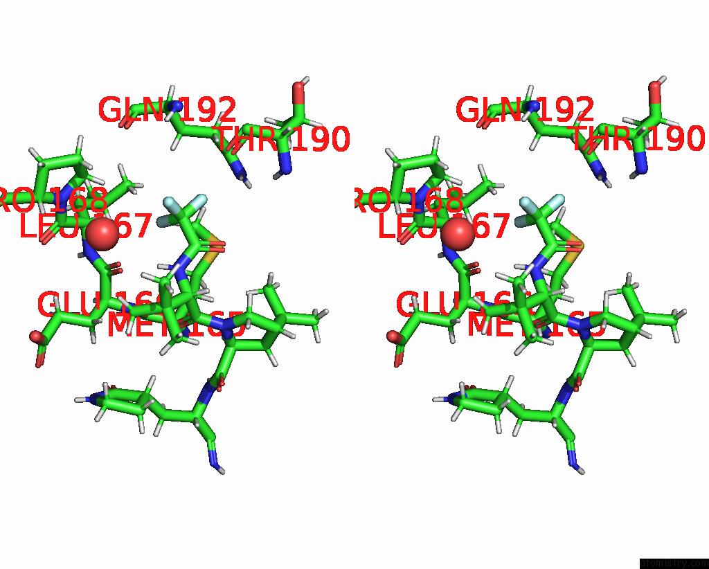
Fluorine binding site 5 out of 6 in 8e25
Go back to
Fluorine binding site 5 out
of 6 in the Crystal Structure of Sars-Cov-2 Main Protease M49I Mutant in Complex with Nirmatrelvir
Mono view
Stereo pair view
Mono view
Stereo pair view
A full contact list of Fluorine with other atoms in the F binding
site number 5 of Crystal Structure of Sars-Cov-2 Main Protease M49I Mutant in Complex with Nirmatrelvir within 5.0Å range:
|
Fluorine binding site 6 out of 6 in 8e25
Go back to
Fluorine binding site 6 out
of 6 in the Crystal Structure of Sars-Cov-2 Main Protease M49I Mutant in Complex with Nirmatrelvir

Mono view
Stereo pair view
Mono view
Stereo pair view
A full contact list of Fluorine with other atoms in the F binding
site number 6 of Crystal Structure of Sars-Cov-2 Main Protease M49I Mutant in Complex with Nirmatrelvir within 5.0Å range:
|
Reference:
G.D.Noske,
A.S.Godoy,
G.Oliva.
Crystal Structure of Sars-Cov-2 Main Protease N142S Mutant in Complex with Nirmatrelvir To Be Published.
Page generated: Wed Jul 16 03:36:45 2025
Last articles
Mn in 9MBLMn in 9MBJ
Mn in 9MBI
Mn in 9MBH
Mn in 9L56
Mn in 9KOQ
Mn in 9KOJ
Mn in 9KQC
Mn in 9JOA
Mn in 9JOB