Fluorine »
PDB 8ez4-8fav »
8f83 »
Fluorine in PDB 8f83: Crystal Structure of Dihydrofolate Reductase (Dhfr) From Mycobacterium Ulcerans AGY99 in Complex with Nadp and Inhibitor MAM846
Enzymatic activity of Crystal Structure of Dihydrofolate Reductase (Dhfr) From Mycobacterium Ulcerans AGY99 in Complex with Nadp and Inhibitor MAM846
All present enzymatic activity of Crystal Structure of Dihydrofolate Reductase (Dhfr) From Mycobacterium Ulcerans AGY99 in Complex with Nadp and Inhibitor MAM846:
1.5.1.3;
1.5.1.3;
Protein crystallography data
The structure of Crystal Structure of Dihydrofolate Reductase (Dhfr) From Mycobacterium Ulcerans AGY99 in Complex with Nadp and Inhibitor MAM846, PDB code: 8f83
was solved by
Seattle Structural Genomics Center For Infectious Disease,
Seattlestructural Genomics Center For Infectious Disease (Ssgcid),
with X-Ray Crystallography technique. A brief refinement statistics is given in the table below:
| Resolution Low / High (Å) | 35.38 / 1.32 |
| Space group | P 1 21 1 |
| Cell size a, b, c (Å), α, β, γ (°) | 29.225, 70.758, 36.067, 90, 111.28, 90 |
| R / Rfree (%) | 15.3 / 18.4 |
Fluorine Binding Sites:
The binding sites of Fluorine atom in the Crystal Structure of Dihydrofolate Reductase (Dhfr) From Mycobacterium Ulcerans AGY99 in Complex with Nadp and Inhibitor MAM846
(pdb code 8f83). This binding sites where shown within
5.0 Angstroms radius around Fluorine atom.
In total 3 binding sites of Fluorine where determined in the Crystal Structure of Dihydrofolate Reductase (Dhfr) From Mycobacterium Ulcerans AGY99 in Complex with Nadp and Inhibitor MAM846, PDB code: 8f83:
Jump to Fluorine binding site number: 1; 2; 3;
In total 3 binding sites of Fluorine where determined in the Crystal Structure of Dihydrofolate Reductase (Dhfr) From Mycobacterium Ulcerans AGY99 in Complex with Nadp and Inhibitor MAM846, PDB code: 8f83:
Jump to Fluorine binding site number: 1; 2; 3;
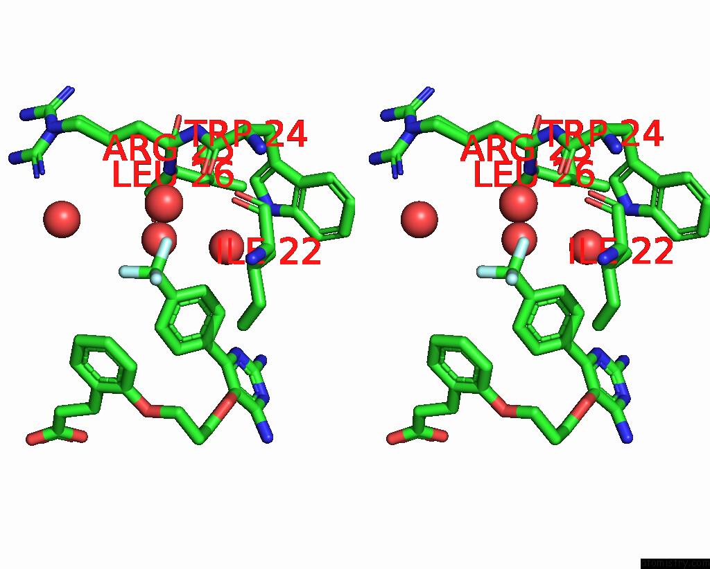
Fluorine binding site 1 out of 3 in 8f83
Go back to
Fluorine binding site 1 out
of 3 in the Crystal Structure of Dihydrofolate Reductase (Dhfr) From Mycobacterium Ulcerans AGY99 in Complex with Nadp and Inhibitor MAM846

Mono view
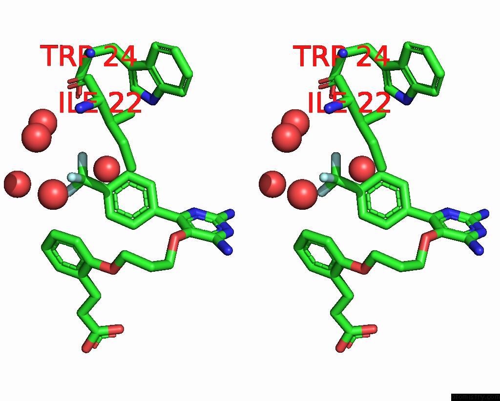
Stereo pair view
Mono view
Stereo pair view
A full contact list of Fluorine with other atoms in the F binding
site number 1 of Crystal Structure of Dihydrofolate Reductase (Dhfr) From Mycobacterium Ulcerans AGY99 in Complex with Nadp and Inhibitor MAM846 within 5.0Å range:
|
Fluorine binding site 2 out of 3 in 8f83
Go back to
Fluorine binding site 2 out
of 3 in the Crystal Structure of Dihydrofolate Reductase (Dhfr) From Mycobacterium Ulcerans AGY99 in Complex with Nadp and Inhibitor MAM846
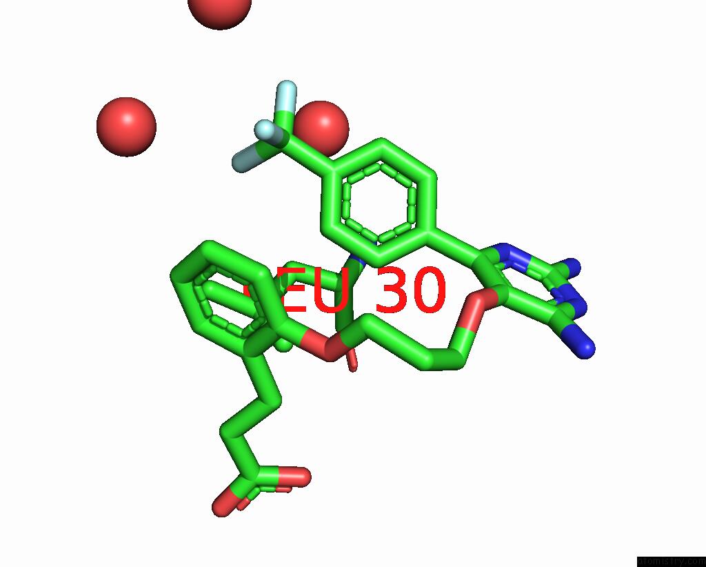
Mono view
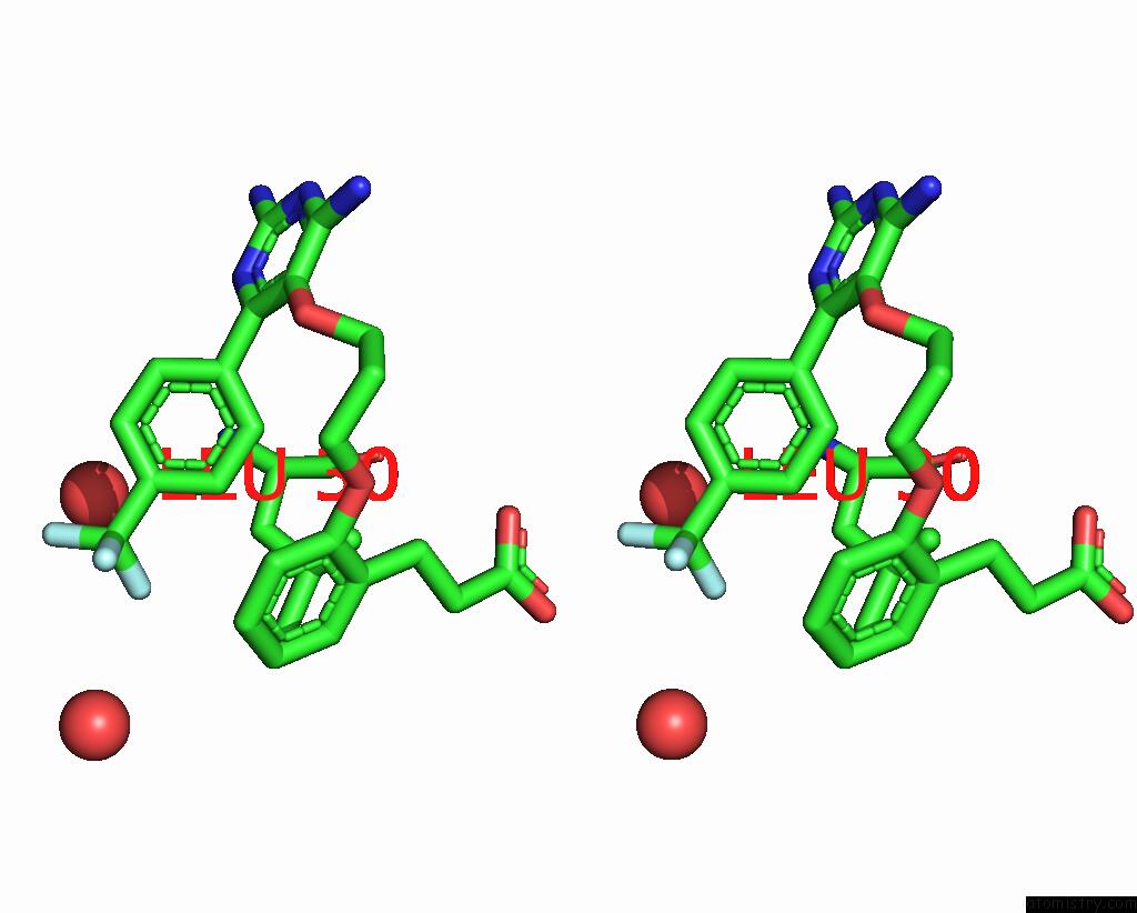
Stereo pair view
Mono view
Stereo pair view
A full contact list of Fluorine with other atoms in the F binding
site number 2 of Crystal Structure of Dihydrofolate Reductase (Dhfr) From Mycobacterium Ulcerans AGY99 in Complex with Nadp and Inhibitor MAM846 within 5.0Å range:
|
Fluorine binding site 3 out of 3 in 8f83
Go back to
Fluorine binding site 3 out
of 3 in the Crystal Structure of Dihydrofolate Reductase (Dhfr) From Mycobacterium Ulcerans AGY99 in Complex with Nadp and Inhibitor MAM846
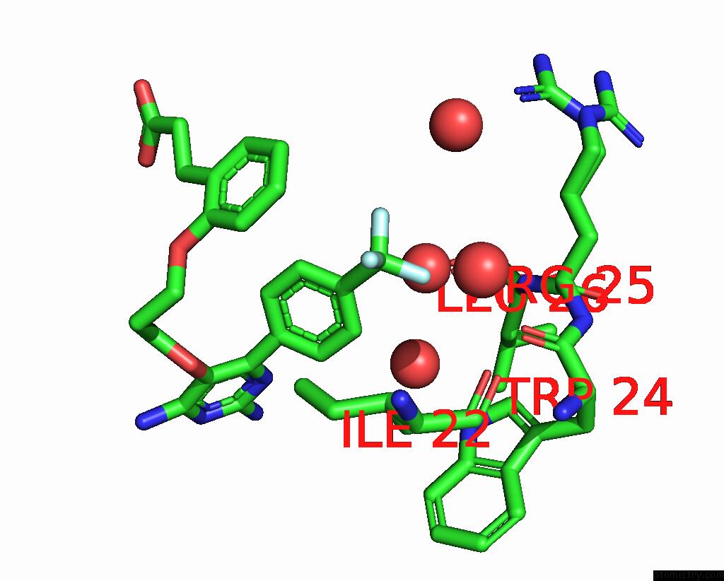
Mono view
Stereo pair view
Mono view
Stereo pair view
A full contact list of Fluorine with other atoms in the F binding
site number 3 of Crystal Structure of Dihydrofolate Reductase (Dhfr) From Mycobacterium Ulcerans AGY99 in Complex with Nadp and Inhibitor MAM846 within 5.0Å range:
|
Reference:
S.Seibold,
K.P.Battaile,
S.Lovell,
B.L.Staker.
Crystal Structure of Dihydrofolate Reductase (Dhfr) From Mycobacterium Ulcerans AGY99 in Complex with Nadp and Inhibitor MAM846 To Be Published.
Page generated: Wed Jul 16 04:11:21 2025
Last articles
Na in 3DDKNa in 3DFH
Na in 3DEB
Na in 3DAV
Na in 3DDR
Na in 3DBO
Na in 3DC7
Na in 3D97
Na in 3DA9
Na in 3D9R