Fluorine »
PDB 8phl-8q0w »
8pie »
Fluorine in PDB 8pie: Crystal Structure of the Human Nucleoside Diphosphate Kinase B Domain in Complex with the Product at-8500 Formed By Catalysis of Compound at-9010
Enzymatic activity of Crystal Structure of the Human Nucleoside Diphosphate Kinase B Domain in Complex with the Product at-8500 Formed By Catalysis of Compound at-9010
All present enzymatic activity of Crystal Structure of the Human Nucleoside Diphosphate Kinase B Domain in Complex with the Product at-8500 Formed By Catalysis of Compound at-9010:
2.7.13.3; 2.7.4.6;
2.7.13.3; 2.7.4.6;
Protein crystallography data
The structure of Crystal Structure of the Human Nucleoside Diphosphate Kinase B Domain in Complex with the Product at-8500 Formed By Catalysis of Compound at-9010, PDB code: 8pie
was solved by
M.Feracci,
A.Chazot,
F.Ferron,
K.Alvarez,
B.Canard,
with X-Ray Crystallography technique. A brief refinement statistics is given in the table below:
| Resolution Low / High (Å) | 49.68 / 1.90 |
| Space group | P 1 21 1 |
| Cell size a, b, c (Å), α, β, γ (°) | 54.19, 120.313, 71.918, 90, 110.15, 90 |
| R / Rfree (%) | 17.2 / 21.3 |
Fluorine Binding Sites:
The binding sites of Fluorine atom in the Crystal Structure of the Human Nucleoside Diphosphate Kinase B Domain in Complex with the Product at-8500 Formed By Catalysis of Compound at-9010
(pdb code 8pie). This binding sites where shown within
5.0 Angstroms radius around Fluorine atom.
In total 6 binding sites of Fluorine where determined in the Crystal Structure of the Human Nucleoside Diphosphate Kinase B Domain in Complex with the Product at-8500 Formed By Catalysis of Compound at-9010, PDB code: 8pie:
Jump to Fluorine binding site number: 1; 2; 3; 4; 5; 6;
In total 6 binding sites of Fluorine where determined in the Crystal Structure of the Human Nucleoside Diphosphate Kinase B Domain in Complex with the Product at-8500 Formed By Catalysis of Compound at-9010, PDB code: 8pie:
Jump to Fluorine binding site number: 1; 2; 3; 4; 5; 6;
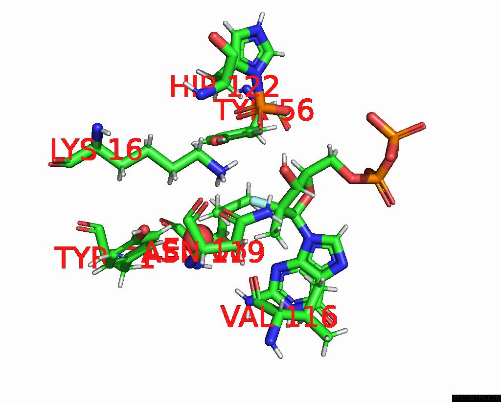
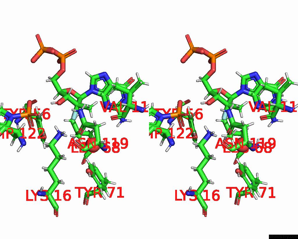
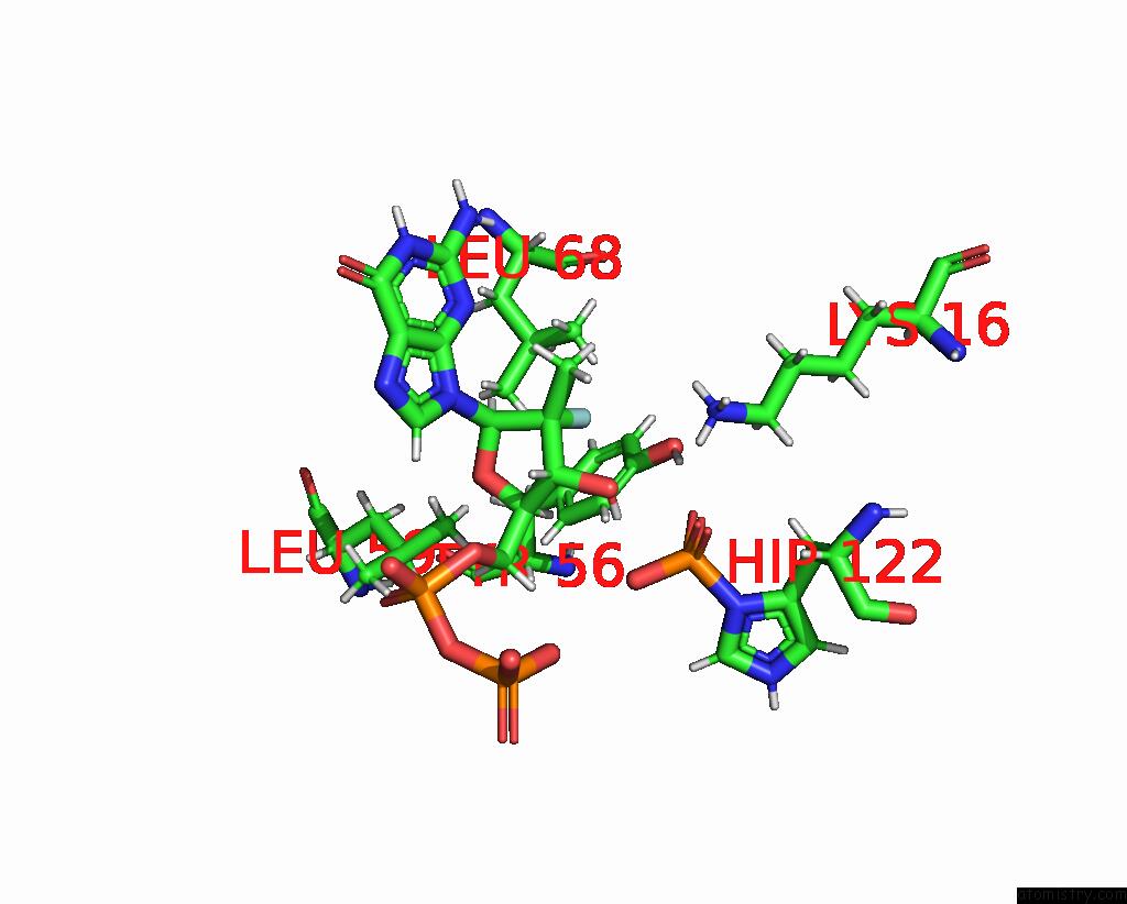
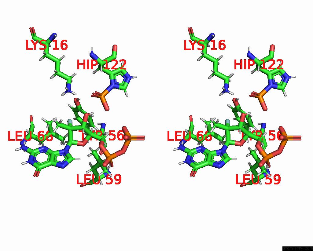
Fluorine binding site 1 out of 6 in 8pie
Go back to
Fluorine binding site 1 out
of 6 in the Crystal Structure of the Human Nucleoside Diphosphate Kinase B Domain in Complex with the Product at-8500 Formed By Catalysis of Compound at-9010
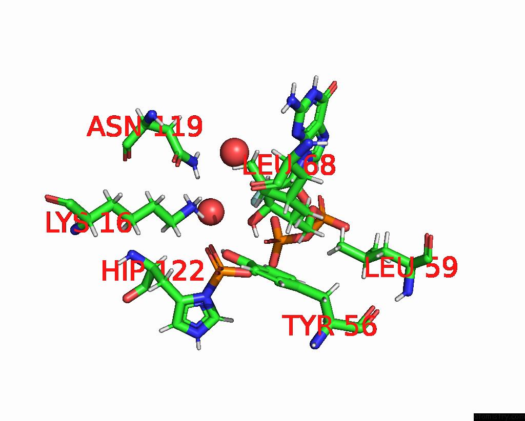
Mono view
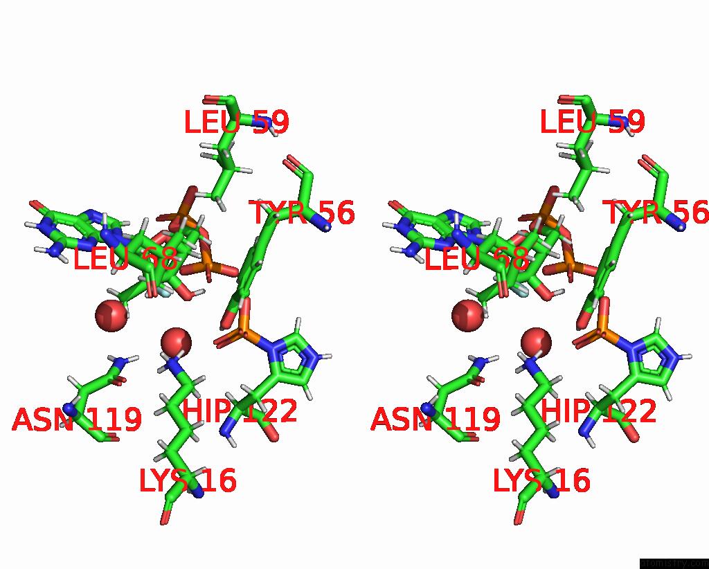
Stereo pair view
Mono view
Stereo pair view
A full contact list of Fluorine with other atoms in the F binding
site number 1 of Crystal Structure of the Human Nucleoside Diphosphate Kinase B Domain in Complex with the Product at-8500 Formed By Catalysis of Compound at-9010 within 5.0Å range:
|
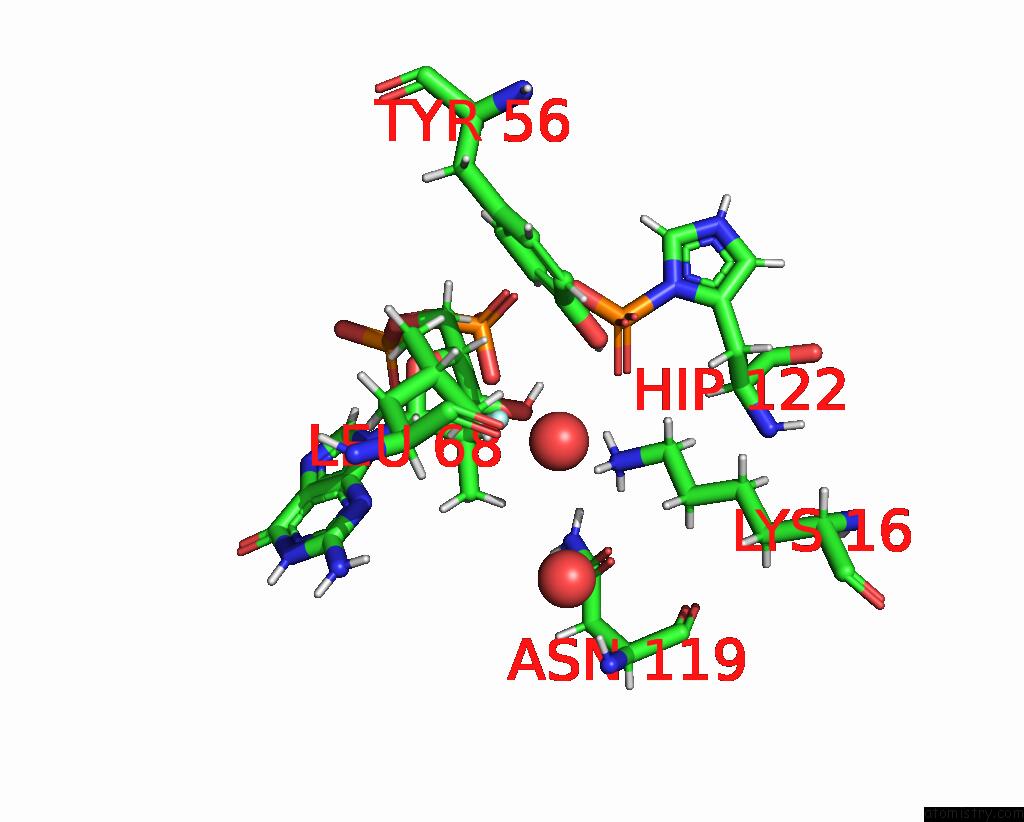
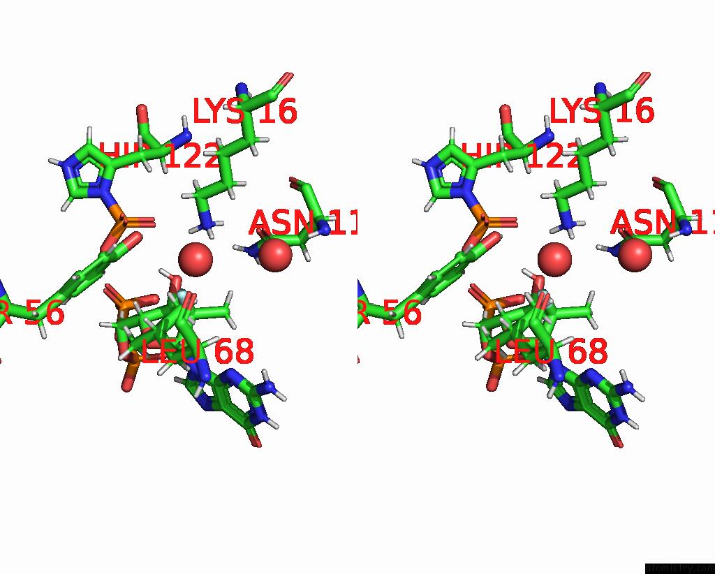
Fluorine binding site 2 out of 6 in 8pie
Go back to
Fluorine binding site 2 out
of 6 in the Crystal Structure of the Human Nucleoside Diphosphate Kinase B Domain in Complex with the Product at-8500 Formed By Catalysis of Compound at-9010
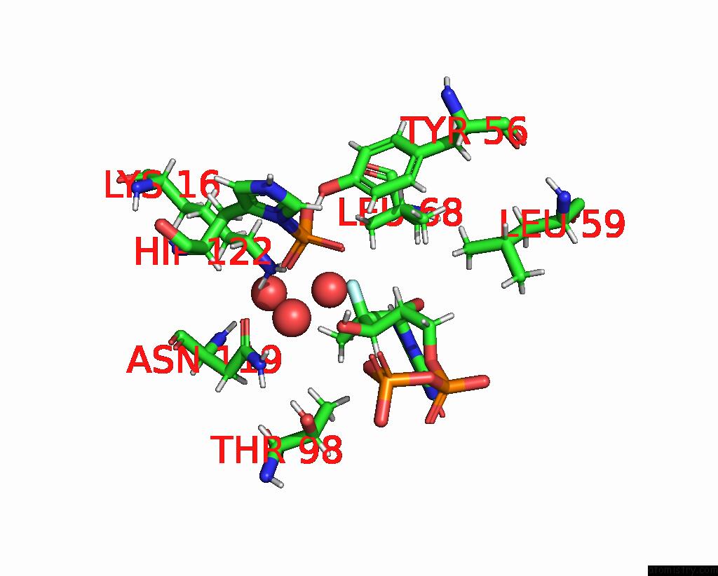
Mono view
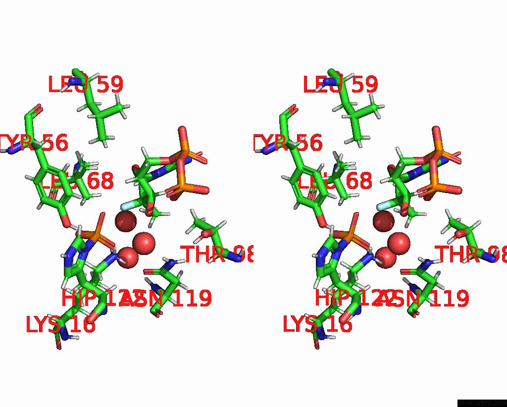
Stereo pair view
Mono view
Stereo pair view
A full contact list of Fluorine with other atoms in the F binding
site number 2 of Crystal Structure of the Human Nucleoside Diphosphate Kinase B Domain in Complex with the Product at-8500 Formed By Catalysis of Compound at-9010 within 5.0Å range:
|
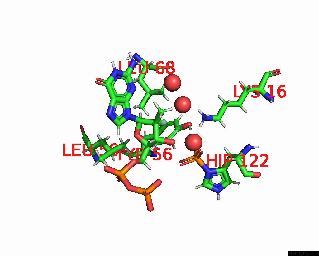
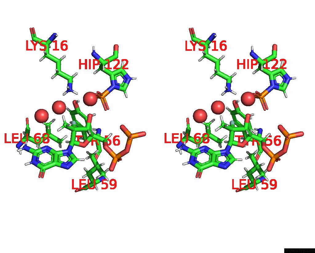
Fluorine binding site 3 out of 6 in 8pie
Go back to
Fluorine binding site 3 out
of 6 in the Crystal Structure of the Human Nucleoside Diphosphate Kinase B Domain in Complex with the Product at-8500 Formed By Catalysis of Compound at-9010
Mono view
Stereo pair view
Mono view
Stereo pair view
A full contact list of Fluorine with other atoms in the F binding
site number 3 of Crystal Structure of the Human Nucleoside Diphosphate Kinase B Domain in Complex with the Product at-8500 Formed By Catalysis of Compound at-9010 within 5.0Å range:
|
Fluorine binding site 4 out of 6 in 8pie
Go back to
Fluorine binding site 4 out
of 6 in the Crystal Structure of the Human Nucleoside Diphosphate Kinase B Domain in Complex with the Product at-8500 Formed By Catalysis of Compound at-9010
Mono view
Stereo pair view
Mono view
Stereo pair view
A full contact list of Fluorine with other atoms in the F binding
site number 4 of Crystal Structure of the Human Nucleoside Diphosphate Kinase B Domain in Complex with the Product at-8500 Formed By Catalysis of Compound at-9010 within 5.0Å range:
|
Fluorine binding site 5 out of 6 in 8pie
Go back to
Fluorine binding site 5 out
of 6 in the Crystal Structure of the Human Nucleoside Diphosphate Kinase B Domain in Complex with the Product at-8500 Formed By Catalysis of Compound at-9010
Mono view
Stereo pair view
Mono view
Stereo pair view
A full contact list of Fluorine with other atoms in the F binding
site number 5 of Crystal Structure of the Human Nucleoside Diphosphate Kinase B Domain in Complex with the Product at-8500 Formed By Catalysis of Compound at-9010 within 5.0Å range:
|
Fluorine binding site 6 out of 6 in 8pie
Go back to
Fluorine binding site 6 out
of 6 in the Crystal Structure of the Human Nucleoside Diphosphate Kinase B Domain in Complex with the Product at-8500 Formed By Catalysis of Compound at-9010
Mono view
Stereo pair view
Mono view
Stereo pair view
A full contact list of Fluorine with other atoms in the F binding
site number 6 of Crystal Structure of the Human Nucleoside Diphosphate Kinase B Domain in Complex with the Product at-8500 Formed By Catalysis of Compound at-9010 within 5.0Å range:
|
Reference:
A.Chazot,
C.Zimberger,
M.Feracci,
A.Moussa,
S.Good,
J.P.Sommadossi,
K.Alvarez,
F.Ferron,
B.Canard.
The Activation Chain of the Broad-Spectrum Antiviral Bemnifosbuvir at Atomic Resolution Plos Biol. 2024.
ISSN: ESSN 1545-7885
DOI: 10.1371/JOURNAL.PBIO.3002743
Page generated: Wed Jul 16 06:39:57 2025
ISSN: ESSN 1545-7885
DOI: 10.1371/JOURNAL.PBIO.3002743
Last articles
Mg in 7TFLMg in 7TFK
Mg in 7TFJ
Mg in 7TFI
Mg in 7TF9
Mg in 7TFE
Mg in 7TDP
Mg in 7TFH
Mg in 7TDB
Mg in 7TE5