Fluorine »
PDB 16pk-1bw7 »
1b0f »
Fluorine in PDB 1b0f: Crystal Structure of Human Neutrophil Elastase with Mdl 101, 146
Enzymatic activity of Crystal Structure of Human Neutrophil Elastase with Mdl 101, 146
All present enzymatic activity of Crystal Structure of Human Neutrophil Elastase with Mdl 101, 146:
3.4.21.37;
3.4.21.37;
Protein crystallography data
The structure of Crystal Structure of Human Neutrophil Elastase with Mdl 101, 146, PDB code: 1b0f
was solved by
H.A.Schreuder,
W.A.Metz,
N.P.Peet,
J.T.Pelton,
C.Tardif,
with X-Ray Crystallography technique. A brief refinement statistics is given in the table below:
| Resolution Low / High (Å) | 8.00 / 3.00 |
| Space group | P 43 2 2 |
| Cell size a, b, c (Å), α, β, γ (°) | 75.800, 75.800, 108.500, 90.00, 90.00, 90.00 |
| R / Rfree (%) | 16 / n/a |
Fluorine Binding Sites:
The binding sites of Fluorine atom in the Crystal Structure of Human Neutrophil Elastase with Mdl 101, 146
(pdb code 1b0f). This binding sites where shown within
5.0 Angstroms radius around Fluorine atom.
In total 5 binding sites of Fluorine where determined in the Crystal Structure of Human Neutrophil Elastase with Mdl 101, 146, PDB code: 1b0f:
Jump to Fluorine binding site number: 1; 2; 3; 4; 5;
In total 5 binding sites of Fluorine where determined in the Crystal Structure of Human Neutrophil Elastase with Mdl 101, 146, PDB code: 1b0f:
Jump to Fluorine binding site number: 1; 2; 3; 4; 5;
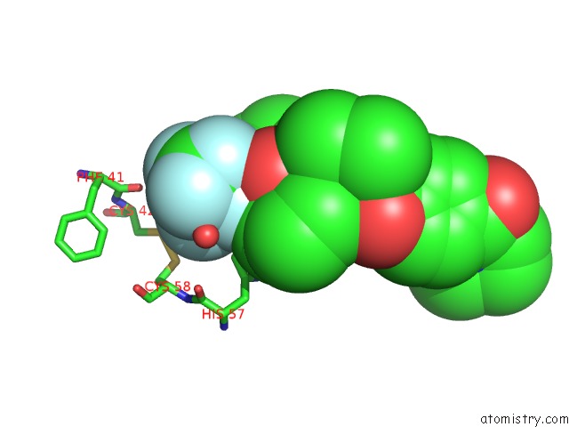
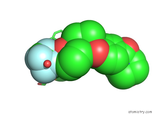
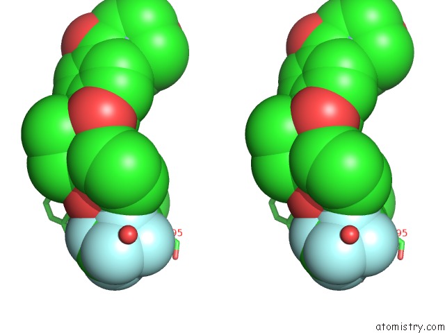
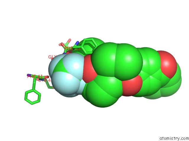
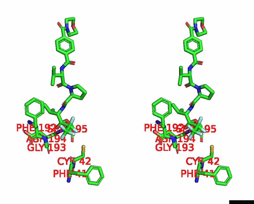
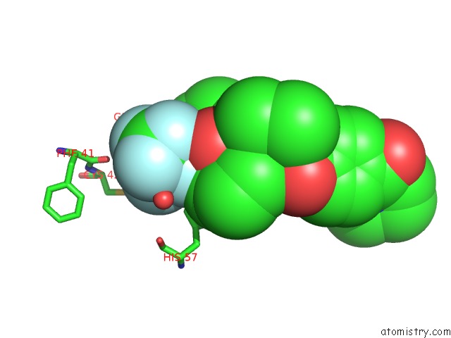
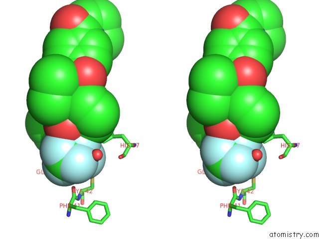
Fluorine binding site 1 out of 5 in 1b0f
Go back to
Fluorine binding site 1 out
of 5 in the Crystal Structure of Human Neutrophil Elastase with Mdl 101, 146
Mono view
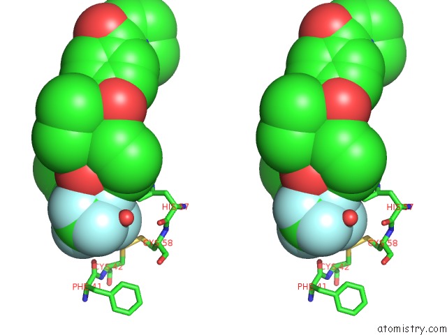
Stereo pair view
Mono view
Stereo pair view
A full contact list of Fluorine with other atoms in the F binding
site number 1 of Crystal Structure of Human Neutrophil Elastase with Mdl 101, 146 within 5.0Å range:
|
Fluorine binding site 2 out of 5 in 1b0f
Go back to
Fluorine binding site 2 out
of 5 in the Crystal Structure of Human Neutrophil Elastase with Mdl 101, 146
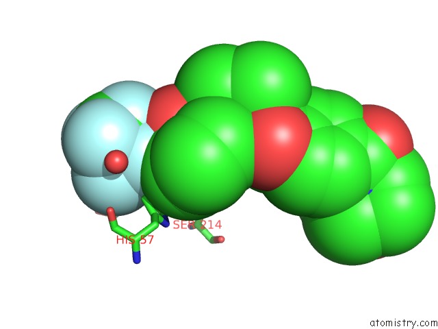
Mono view
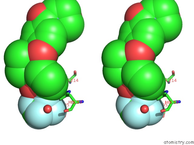
Stereo pair view
Mono view
Stereo pair view
A full contact list of Fluorine with other atoms in the F binding
site number 2 of Crystal Structure of Human Neutrophil Elastase with Mdl 101, 146 within 5.0Å range:
|
Fluorine binding site 3 out of 5 in 1b0f
Go back to
Fluorine binding site 3 out
of 5 in the Crystal Structure of Human Neutrophil Elastase with Mdl 101, 146
Mono view
Stereo pair view
Mono view
Stereo pair view
A full contact list of Fluorine with other atoms in the F binding
site number 3 of Crystal Structure of Human Neutrophil Elastase with Mdl 101, 146 within 5.0Å range:
|
Fluorine binding site 4 out of 5 in 1b0f
Go back to
Fluorine binding site 4 out
of 5 in the Crystal Structure of Human Neutrophil Elastase with Mdl 101, 146
Mono view
Stereo pair view
Mono view
Stereo pair view
A full contact list of Fluorine with other atoms in the F binding
site number 4 of Crystal Structure of Human Neutrophil Elastase with Mdl 101, 146 within 5.0Å range:
|
Fluorine binding site 5 out of 5 in 1b0f
Go back to
Fluorine binding site 5 out
of 5 in the Crystal Structure of Human Neutrophil Elastase with Mdl 101, 146
Mono view
Stereo pair view
Mono view
Stereo pair view
A full contact list of Fluorine with other atoms in the F binding
site number 5 of Crystal Structure of Human Neutrophil Elastase with Mdl 101, 146 within 5.0Å range:
|
Reference:
R.J.Cregge,
S.L.Durham,
R.A.Farr,
S.L.Gallion,
C.M.Hare,
R.V.Hoffman,
M.J.Janusz,
H.O.Kim,
J.R.Koehl,
S.Mehdi,
W.A.Metz,
N.P.Peet,
J.T.Pelton,
H.A.Schreuder,
S.Sunder,
C.Tardif.
Inhibition of Human Neutrophil Elastase. 4. Design, Synthesis, X-Ray Crystallographic Analysis, and Structure-Activity Relationships For A Series of P2-Modified, Orally Active Peptidyl Pentafluoroethyl Ketones. J.Med.Chem. V. 41 2461 1998.
ISSN: ISSN 0022-2623
PubMed: 9651152
DOI: 10.1021/JM970812E
Page generated: Wed Jul 31 10:50:17 2024
ISSN: ISSN 0022-2623
PubMed: 9651152
DOI: 10.1021/JM970812E
Last articles
Zn in 9J0NZn in 9J0O
Zn in 9J0P
Zn in 9FJX
Zn in 9EKB
Zn in 9C0F
Zn in 9CAH
Zn in 9CH0
Zn in 9CH3
Zn in 9CH1