Fluorine »
PDB 1bwf-1dvy »
1c0l »
Fluorine in PDB 1c0l: D-Amino Acid Oxidase: Structure of Substrate Complexes at Very High Resolution Reveal the Chemical Reacttion Mechanism of Flavin Dehydrogenation
Protein crystallography data
The structure of D-Amino Acid Oxidase: Structure of Substrate Complexes at Very High Resolution Reveal the Chemical Reacttion Mechanism of Flavin Dehydrogenation, PDB code: 1c0l
was solved by
S.Umhau,
G.Molla,
K.Diederichs,
M.S.Pilone,
S.Ghisla,
W.Welte,
with X-Ray Crystallography technique. A brief refinement statistics is given in the table below:
| Resolution Low / High (Å) | 100.00 / 1.73 |
| Space group | I 4 2 2 |
| Cell size a, b, c (Å), α, β, γ (°) | 120.355, 120.355, 136.625, 90.00, 90.00, 90.00 |
| R / Rfree (%) | 16.2 / 20.4 |
Fluorine Binding Sites:
The binding sites of Fluorine atom in the D-Amino Acid Oxidase: Structure of Substrate Complexes at Very High Resolution Reveal the Chemical Reacttion Mechanism of Flavin Dehydrogenation
(pdb code 1c0l). This binding sites where shown within
5.0 Angstroms radius around Fluorine atom.
In total 3 binding sites of Fluorine where determined in the D-Amino Acid Oxidase: Structure of Substrate Complexes at Very High Resolution Reveal the Chemical Reacttion Mechanism of Flavin Dehydrogenation, PDB code: 1c0l:
Jump to Fluorine binding site number: 1; 2; 3;
In total 3 binding sites of Fluorine where determined in the D-Amino Acid Oxidase: Structure of Substrate Complexes at Very High Resolution Reveal the Chemical Reacttion Mechanism of Flavin Dehydrogenation, PDB code: 1c0l:
Jump to Fluorine binding site number: 1; 2; 3;
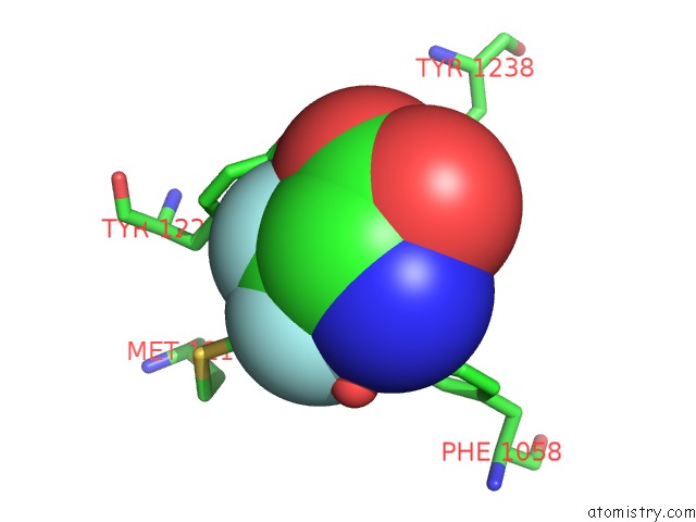
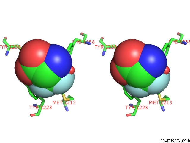
Fluorine binding site 1 out of 3 in 1c0l
Go back to
Fluorine binding site 1 out
of 3 in the D-Amino Acid Oxidase: Structure of Substrate Complexes at Very High Resolution Reveal the Chemical Reacttion Mechanism of Flavin Dehydrogenation
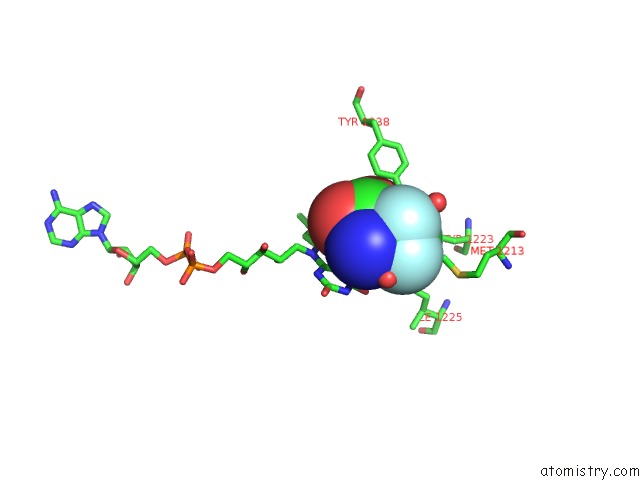
Mono view
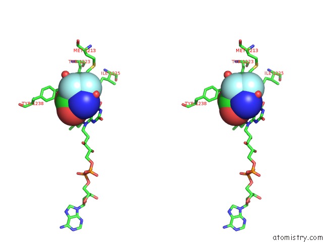
Stereo pair view
Mono view
Stereo pair view
A full contact list of Fluorine with other atoms in the F binding
site number 1 of D-Amino Acid Oxidase: Structure of Substrate Complexes at Very High Resolution Reveal the Chemical Reacttion Mechanism of Flavin Dehydrogenation within 5.0Å range:
|
Fluorine binding site 2 out of 3 in 1c0l
Go back to
Fluorine binding site 2 out
of 3 in the D-Amino Acid Oxidase: Structure of Substrate Complexes at Very High Resolution Reveal the Chemical Reacttion Mechanism of Flavin Dehydrogenation
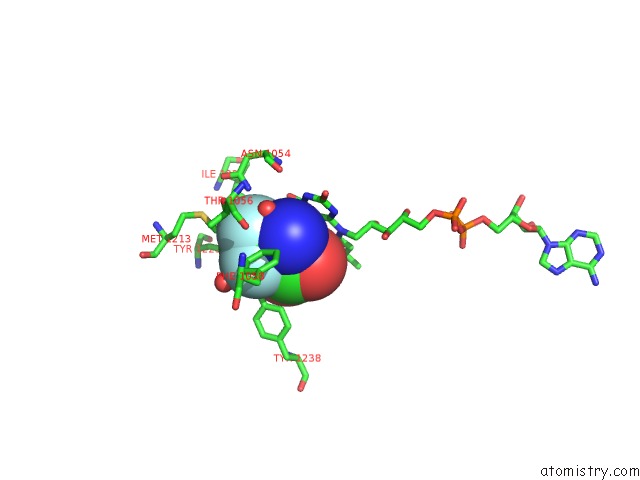
Mono view
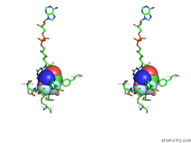
Stereo pair view
Mono view
Stereo pair view
A full contact list of Fluorine with other atoms in the F binding
site number 2 of D-Amino Acid Oxidase: Structure of Substrate Complexes at Very High Resolution Reveal the Chemical Reacttion Mechanism of Flavin Dehydrogenation within 5.0Å range:
|
Fluorine binding site 3 out of 3 in 1c0l
Go back to
Fluorine binding site 3 out
of 3 in the D-Amino Acid Oxidase: Structure of Substrate Complexes at Very High Resolution Reveal the Chemical Reacttion Mechanism of Flavin Dehydrogenation
Mono view
Stereo pair view
Mono view
Stereo pair view
A full contact list of Fluorine with other atoms in the F binding
site number 3 of D-Amino Acid Oxidase: Structure of Substrate Complexes at Very High Resolution Reveal the Chemical Reacttion Mechanism of Flavin Dehydrogenation within 5.0Å range:
|
Reference:
S.Umhau,
L.Pollegioni,
G.Molla,
K.Diederichs,
W.Welte,
M.S.Pilone,
S.Ghisla.
The X-Ray Structure of D-Amino Acid Oxidase at Very High Resolution Identifies the Chemical Mechanism of Flavin-Dependent Substrate Dehydrogenation. Proc.Natl.Acad.Sci.Usa V. 97 12463 2000.
ISSN: ISSN 0027-8424
PubMed: 11070076
DOI: 10.1073/PNAS.97.23.12463
Page generated: Wed Jul 31 10:59:41 2024
ISSN: ISSN 0027-8424
PubMed: 11070076
DOI: 10.1073/PNAS.97.23.12463
Last articles
Zn in 9MJ5Zn in 9HNW
Zn in 9G0L
Zn in 9FNE
Zn in 9DZN
Zn in 9E0I
Zn in 9D32
Zn in 9DAK
Zn in 8ZXC
Zn in 8ZUF