Fluorine »
PDB 1bwf-1dvy »
1c22 »
Fluorine in PDB 1c22: E. Coli Methionine Aminopeptidase: Trifluoromethionine Complex
Enzymatic activity of E. Coli Methionine Aminopeptidase: Trifluoromethionine Complex
All present enzymatic activity of E. Coli Methionine Aminopeptidase: Trifluoromethionine Complex:
3.4.11.18;
3.4.11.18;
Protein crystallography data
The structure of E. Coli Methionine Aminopeptidase: Trifluoromethionine Complex, PDB code: 1c22
was solved by
W.T.Lowther,
Y.Zhang,
P.B.Sampson,
J.F.Honek,
B.W.Matthews,
with X-Ray Crystallography technique. A brief refinement statistics is given in the table below:
| Resolution Low / High (Å) | 27.10 / 1.75 |
| Space group | P 1 21 1 |
| Cell size a, b, c (Å), α, β, γ (°) | 39.128, 67.604, 48.826, 90.00, 111.05, 90.00 |
| R / Rfree (%) | n/a / n/a |
Other elements in 1c22:
The structure of E. Coli Methionine Aminopeptidase: Trifluoromethionine Complex also contains other interesting chemical elements:
| Cobalt | (Co) | 2 atoms |
| Sodium | (Na) | 1 atom |
Fluorine Binding Sites:
The binding sites of Fluorine atom in the E. Coli Methionine Aminopeptidase: Trifluoromethionine Complex
(pdb code 1c22). This binding sites where shown within
5.0 Angstroms radius around Fluorine atom.
In total 3 binding sites of Fluorine where determined in the E. Coli Methionine Aminopeptidase: Trifluoromethionine Complex, PDB code: 1c22:
Jump to Fluorine binding site number: 1; 2; 3;
In total 3 binding sites of Fluorine where determined in the E. Coli Methionine Aminopeptidase: Trifluoromethionine Complex, PDB code: 1c22:
Jump to Fluorine binding site number: 1; 2; 3;
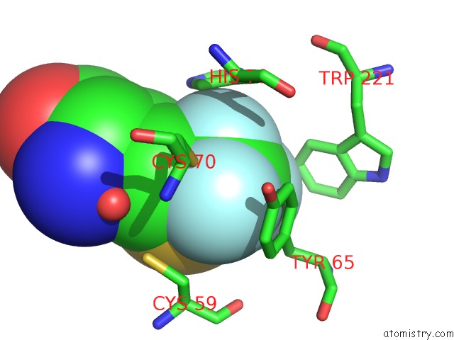
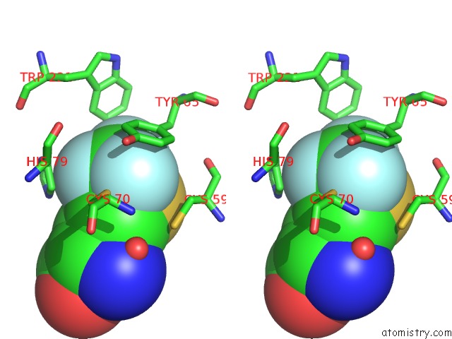
Fluorine binding site 1 out of 3 in 1c22
Go back to
Fluorine binding site 1 out
of 3 in the E. Coli Methionine Aminopeptidase: Trifluoromethionine Complex
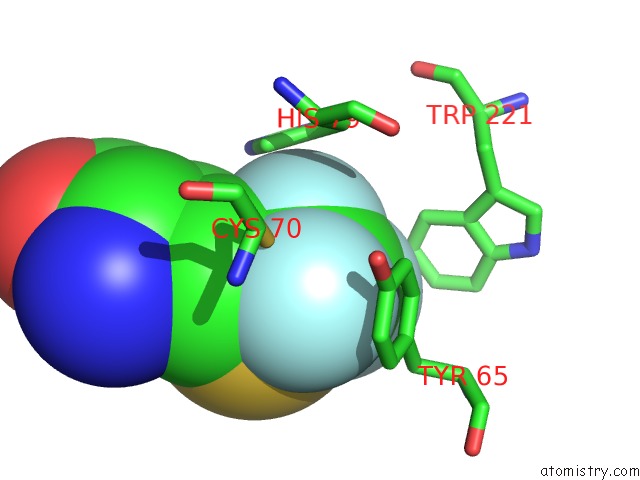
Mono view
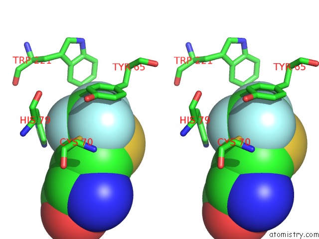
Stereo pair view
Mono view
Stereo pair view
A full contact list of Fluorine with other atoms in the F binding
site number 1 of E. Coli Methionine Aminopeptidase: Trifluoromethionine Complex within 5.0Å range:
|
Fluorine binding site 2 out of 3 in 1c22
Go back to
Fluorine binding site 2 out
of 3 in the E. Coli Methionine Aminopeptidase: Trifluoromethionine Complex
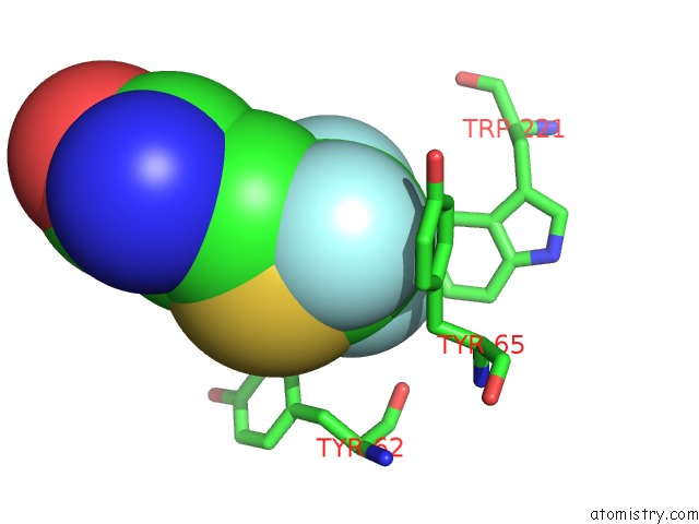
Mono view
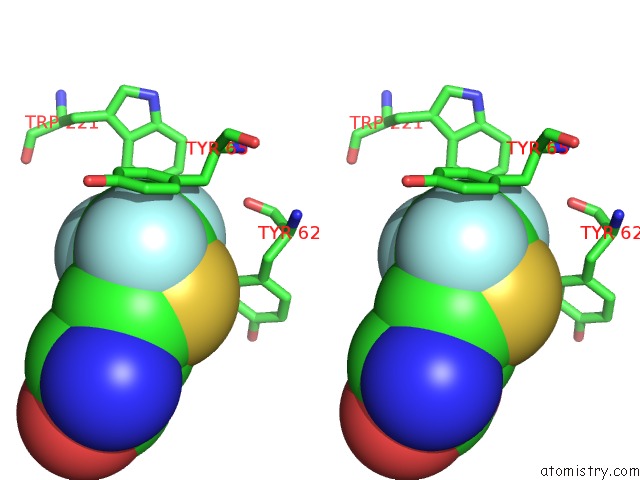
Stereo pair view
Mono view
Stereo pair view
A full contact list of Fluorine with other atoms in the F binding
site number 2 of E. Coli Methionine Aminopeptidase: Trifluoromethionine Complex within 5.0Å range:
|
Fluorine binding site 3 out of 3 in 1c22
Go back to
Fluorine binding site 3 out
of 3 in the E. Coli Methionine Aminopeptidase: Trifluoromethionine Complex
Mono view
Stereo pair view
Mono view
Stereo pair view
A full contact list of Fluorine with other atoms in the F binding
site number 3 of E. Coli Methionine Aminopeptidase: Trifluoromethionine Complex within 5.0Å range:
|
Reference:
W.T.Lowther,
Y.Zhang,
P.B.Sampson,
J.F.Honek,
B.W.Matthews.
Insights Into the Mechanism of Escherichia Coli Methionine Aminopeptidase From the Structural Analysis of Reaction Products and Phosphorus-Based Transition-State Analogues. Biochemistry V. 38 14810 1999.
ISSN: ISSN 0006-2960
PubMed: 10555963
DOI: 10.1021/BI991711G
Page generated: Mon Jul 14 10:28:41 2025
ISSN: ISSN 0006-2960
PubMed: 10555963
DOI: 10.1021/BI991711G
Last articles
Fe in 2YXOFe in 2YRS
Fe in 2YXC
Fe in 2YNM
Fe in 2YVJ
Fe in 2YP1
Fe in 2YU2
Fe in 2YU1
Fe in 2YQB
Fe in 2YOO