Fluorine »
PDB 1dvz-1fk9 »
1e0v »
Fluorine in PDB 1e0v: Xylanase 10A From Sreptomyces Lividans. Cellobiosyl-Enzyme Intermediate at 1.7 A
Enzymatic activity of Xylanase 10A From Sreptomyces Lividans. Cellobiosyl-Enzyme Intermediate at 1.7 A
All present enzymatic activity of Xylanase 10A From Sreptomyces Lividans. Cellobiosyl-Enzyme Intermediate at 1.7 A:
3.2.1.8;
3.2.1.8;
Protein crystallography data
The structure of Xylanase 10A From Sreptomyces Lividans. Cellobiosyl-Enzyme Intermediate at 1.7 A, PDB code: 1e0v
was solved by
V.Ducros,
S.J.Charnock,
U.Derewenda,
Z.S.Derewenda,
Z.Dauter,
C.Dupont,
F.Shareck,
R.Morosoli,
D.Kluepfel,
G.J.Davies,
with X-Ray Crystallography technique. A brief refinement statistics is given in the table below:
| Resolution Low / High (Å) | 15 / 1.7 |
| Space group | P 21 21 21 |
| Cell size a, b, c (Å), α, β, γ (°) | 67.870, 46.270, 87.070, 90.00, 90.00, 90.00 |
| R / Rfree (%) | 15.8 / 19.9 |
Fluorine Binding Sites:
The binding sites of Fluorine atom in the Xylanase 10A From Sreptomyces Lividans. Cellobiosyl-Enzyme Intermediate at 1.7 A
(pdb code 1e0v). This binding sites where shown within
5.0 Angstroms radius around Fluorine atom.
In total only one binding site of Fluorine was determined in the Xylanase 10A From Sreptomyces Lividans. Cellobiosyl-Enzyme Intermediate at 1.7 A, PDB code: 1e0v:
In total only one binding site of Fluorine was determined in the Xylanase 10A From Sreptomyces Lividans. Cellobiosyl-Enzyme Intermediate at 1.7 A, PDB code: 1e0v:
Fluorine binding site 1 out of 1 in 1e0v
Go back to
Fluorine binding site 1 out
of 1 in the Xylanase 10A From Sreptomyces Lividans. Cellobiosyl-Enzyme Intermediate at 1.7 A
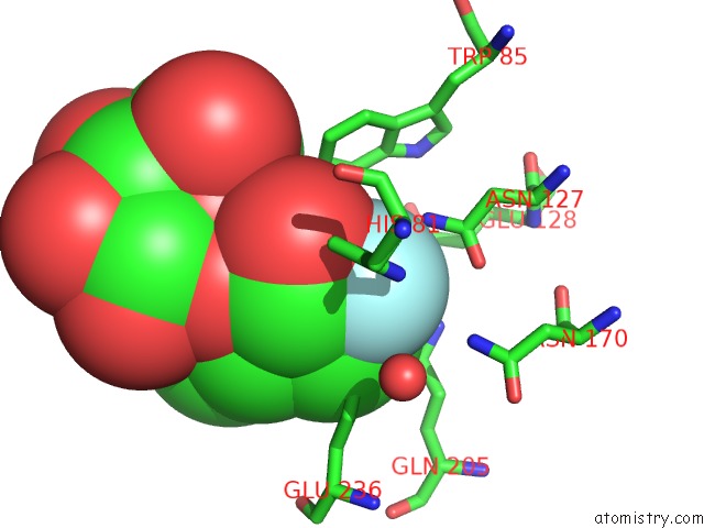
Mono view
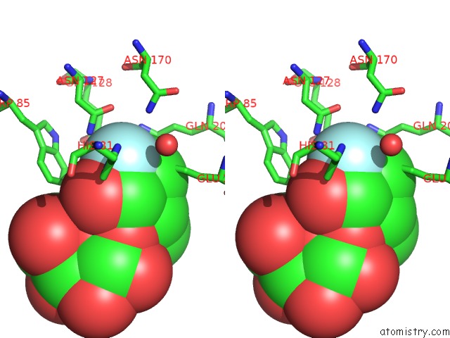
Stereo pair view
Mono view
Stereo pair view
A full contact list of Fluorine with other atoms in the F binding
site number 1 of Xylanase 10A From Sreptomyces Lividans. Cellobiosyl-Enzyme Intermediate at 1.7 A within 5.0Å range:
|
Reference:
V.Ducros,
S.J.Charnock,
U.Derewenda,
Z.S.Derewenda,
Z.Dauter,
C.Dupont,
F.Shareck,
R.Morosoli,
D.Kluepfel,
G.J.Davies.
Substrate Specificity in Glycoside Hydrolase Family 10. Structural and Kinetic Analysis of the Streptomyces Lividans Xylanase 10A J.Biol.Chem. V. 275 23020 2000.
ISSN: ISSN 0021-9258
PubMed: 10930426
DOI: 10.1074/JBC.M000129200
Page generated: Wed Jul 31 11:07:40 2024
ISSN: ISSN 0021-9258
PubMed: 10930426
DOI: 10.1074/JBC.M000129200
Last articles
Zn in 9J0NZn in 9J0O
Zn in 9J0P
Zn in 9FJX
Zn in 9EKB
Zn in 9C0F
Zn in 9CAH
Zn in 9CH0
Zn in 9CH3
Zn in 9CH1