Fluorine »
PDB 1jdj-1mkd »
1l3r »
Fluorine in PDB 1l3r: Crystal Structure of A Transition State Mimic of the Catalytic Subunit of Camp-Dependent Protein Kinase
Enzymatic activity of Crystal Structure of A Transition State Mimic of the Catalytic Subunit of Camp-Dependent Protein Kinase
All present enzymatic activity of Crystal Structure of A Transition State Mimic of the Catalytic Subunit of Camp-Dependent Protein Kinase:
2.7.1.37;
2.7.1.37;
Protein crystallography data
The structure of Crystal Structure of A Transition State Mimic of the Catalytic Subunit of Camp-Dependent Protein Kinase, PDB code: 1l3r
was solved by
Madhusudan,
P.Akamine,
N.-H.Xuong,
S.S.Taylor,
with X-Ray Crystallography technique. A brief refinement statistics is given in the table below:
| Resolution Low / High (Å) | 35.00 / 2.00 |
| Space group | P 21 21 21 |
| Cell size a, b, c (Å), α, β, γ (°) | 72.680, 75.700, 80.550, 90.00, 90.00, 90.00 |
| R / Rfree (%) | 20.5 / 23.2 |
Other elements in 1l3r:
The structure of Crystal Structure of A Transition State Mimic of the Catalytic Subunit of Camp-Dependent Protein Kinase also contains other interesting chemical elements:
| Magnesium | (Mg) | 2 atoms |
| Aluminium | (Al) | 1 atom |
Fluorine Binding Sites:
The binding sites of Fluorine atom in the Crystal Structure of A Transition State Mimic of the Catalytic Subunit of Camp-Dependent Protein Kinase
(pdb code 1l3r). This binding sites where shown within
5.0 Angstroms radius around Fluorine atom.
In total 3 binding sites of Fluorine where determined in the Crystal Structure of A Transition State Mimic of the Catalytic Subunit of Camp-Dependent Protein Kinase, PDB code: 1l3r:
Jump to Fluorine binding site number: 1; 2; 3;
In total 3 binding sites of Fluorine where determined in the Crystal Structure of A Transition State Mimic of the Catalytic Subunit of Camp-Dependent Protein Kinase, PDB code: 1l3r:
Jump to Fluorine binding site number: 1; 2; 3;
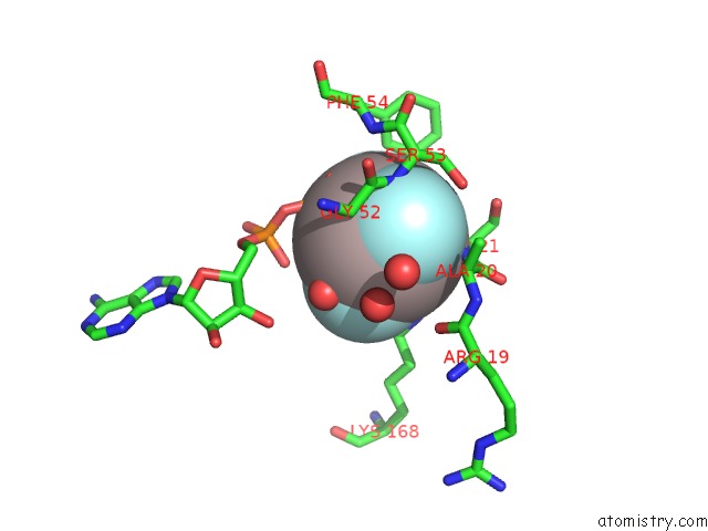
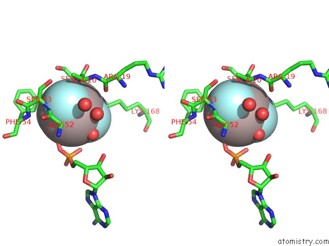
Fluorine binding site 1 out of 3 in 1l3r
Go back to
Fluorine binding site 1 out
of 3 in the Crystal Structure of A Transition State Mimic of the Catalytic Subunit of Camp-Dependent Protein Kinase
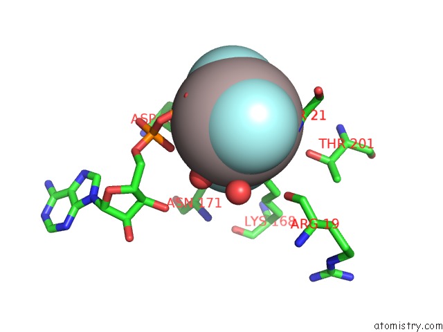
Mono view
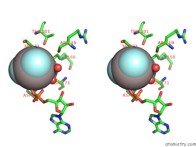
Stereo pair view
Mono view
Stereo pair view
A full contact list of Fluorine with other atoms in the F binding
site number 1 of Crystal Structure of A Transition State Mimic of the Catalytic Subunit of Camp-Dependent Protein Kinase within 5.0Å range:
|
Fluorine binding site 2 out of 3 in 1l3r
Go back to
Fluorine binding site 2 out
of 3 in the Crystal Structure of A Transition State Mimic of the Catalytic Subunit of Camp-Dependent Protein Kinase
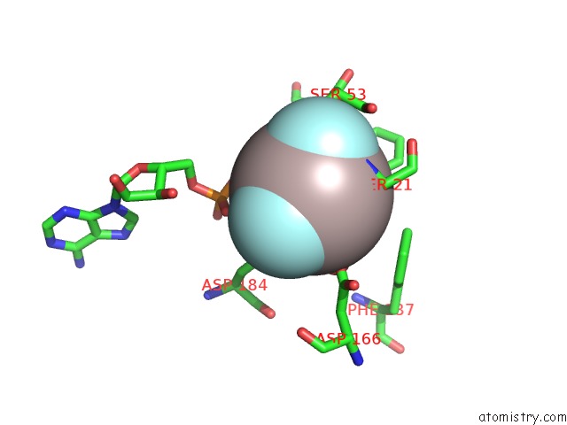
Mono view
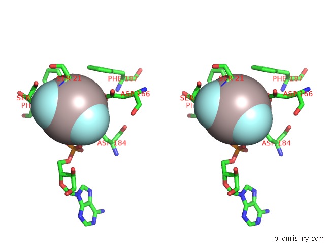
Stereo pair view
Mono view
Stereo pair view
A full contact list of Fluorine with other atoms in the F binding
site number 2 of Crystal Structure of A Transition State Mimic of the Catalytic Subunit of Camp-Dependent Protein Kinase within 5.0Å range:
|
Fluorine binding site 3 out of 3 in 1l3r
Go back to
Fluorine binding site 3 out
of 3 in the Crystal Structure of A Transition State Mimic of the Catalytic Subunit of Camp-Dependent Protein Kinase
Mono view
Stereo pair view
Mono view
Stereo pair view
A full contact list of Fluorine with other atoms in the F binding
site number 3 of Crystal Structure of A Transition State Mimic of the Catalytic Subunit of Camp-Dependent Protein Kinase within 5.0Å range:
|
Reference:
Madhusudan,
P.Akamine,
N.H.Xuong,
S.S.Taylor.
Crystal Structure of A Transition State Mimic of the Catalytic Subunit of Camp-Dependent Protein Kinase. Nat.Struct.Biol. V. 9 273 2002.
ISSN: ISSN 1072-8368
PubMed: 11896404
DOI: 10.1038/NSB780
Page generated: Wed Jul 31 11:46:33 2024
ISSN: ISSN 1072-8368
PubMed: 11896404
DOI: 10.1038/NSB780
Last articles
Zn in 9J0NZn in 9J0O
Zn in 9J0P
Zn in 9FJX
Zn in 9EKB
Zn in 9C0F
Zn in 9CAH
Zn in 9CH0
Zn in 9CH3
Zn in 9CH1