Fluorine »
PDB 1jdj-1mkd »
1lgw »
Fluorine in PDB 1lgw: T4 Lysozyme Mutant L99A/M102Q Bound By 2-Fluoroaniline
Enzymatic activity of T4 Lysozyme Mutant L99A/M102Q Bound By 2-Fluoroaniline
All present enzymatic activity of T4 Lysozyme Mutant L99A/M102Q Bound By 2-Fluoroaniline:
3.2.1.17;
3.2.1.17;
Protein crystallography data
The structure of T4 Lysozyme Mutant L99A/M102Q Bound By 2-Fluoroaniline, PDB code: 1lgw
was solved by
B.Q.Wei,
W.A.Baase,
L.H.Weaver,
B.W.Matthews,
B.K.Shoichet,
with X-Ray Crystallography technique. A brief refinement statistics is given in the table below:
| Resolution Low / High (Å) | 15.00 / 1.85 |
| Space group | P 32 2 1 |
| Cell size a, b, c (Å), α, β, γ (°) | 60.800, 60.800, 97.740, 90.00, 90.00, 120.00 |
| R / Rfree (%) | n/a / n/a |
Other elements in 1lgw:
The structure of T4 Lysozyme Mutant L99A/M102Q Bound By 2-Fluoroaniline also contains other interesting chemical elements:
| Chlorine | (Cl) | 2 atoms |
Fluorine Binding Sites:
The binding sites of Fluorine atom in the T4 Lysozyme Mutant L99A/M102Q Bound By 2-Fluoroaniline
(pdb code 1lgw). This binding sites where shown within
5.0 Angstroms radius around Fluorine atom.
In total only one binding site of Fluorine was determined in the T4 Lysozyme Mutant L99A/M102Q Bound By 2-Fluoroaniline, PDB code: 1lgw:
In total only one binding site of Fluorine was determined in the T4 Lysozyme Mutant L99A/M102Q Bound By 2-Fluoroaniline, PDB code: 1lgw:
Fluorine binding site 1 out of 1 in 1lgw
Go back to
Fluorine binding site 1 out
of 1 in the T4 Lysozyme Mutant L99A/M102Q Bound By 2-Fluoroaniline
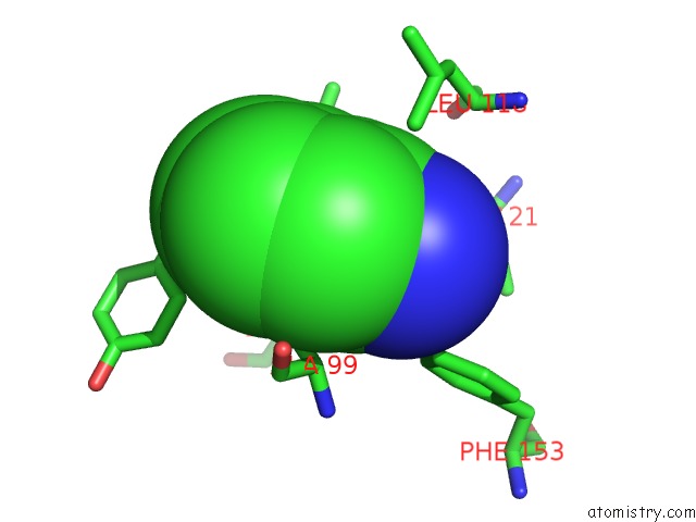
Mono view
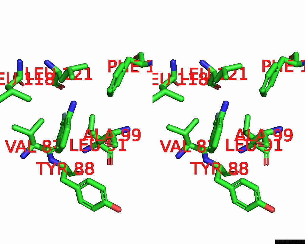
Stereo pair view
Mono view
Stereo pair view
A full contact list of Fluorine with other atoms in the F binding
site number 1 of T4 Lysozyme Mutant L99A/M102Q Bound By 2-Fluoroaniline within 5.0Å range:
|
Reference:
B.Q.Wei,
W.A.Baase,
L.H.Weaver,
B.W.Matthews,
B.K.Shoichet.
A Model Binding Site For Testing Scoring Functions in Molecular Docking J.Mol.Biol. V. 322 339 2002.
ISSN: ISSN 0022-2836
PubMed: 12217695
DOI: 10.1016/S0022-2836(02)00777-5
Page generated: Wed Jul 31 11:47:22 2024
ISSN: ISSN 0022-2836
PubMed: 12217695
DOI: 10.1016/S0022-2836(02)00777-5
Last articles
Zn in 9J0NZn in 9J0O
Zn in 9J0P
Zn in 9FJX
Zn in 9EKB
Zn in 9C0F
Zn in 9CAH
Zn in 9CH0
Zn in 9CH3
Zn in 9CH1