Fluorine »
PDB 1jdj-1mkd »
1mfi »
Fluorine in PDB 1mfi: Crystal Structure of Macrophage Migration Inhibitory Factor Complexed with (E)-2-Fluoro-P-Hydroxycinnamate
Protein crystallography data
The structure of Crystal Structure of Macrophage Migration Inhibitory Factor Complexed with (E)-2-Fluoro-P-Hydroxycinnamate, PDB code: 1mfi
was solved by
A.B.Taylor,
W.H.Johnson Jr.,
R.M.Czerwinski,
C.P.Whitman,
M.L.Hackert,
with X-Ray Crystallography technique. A brief refinement statistics is given in the table below:
| Resolution Low / High (Å) | 50.00 / 1.80 |
| Space group | H 3 |
| Cell size a, b, c (Å), α, β, γ (°) | 99.530, 99.530, 105.690, 90.00, 90.00, 120.00 |
| R / Rfree (%) | 17.8 / 22.7 |
Fluorine Binding Sites:
The binding sites of Fluorine atom in the Crystal Structure of Macrophage Migration Inhibitory Factor Complexed with (E)-2-Fluoro-P-Hydroxycinnamate
(pdb code 1mfi). This binding sites where shown within
5.0 Angstroms radius around Fluorine atom.
In total 3 binding sites of Fluorine where determined in the Crystal Structure of Macrophage Migration Inhibitory Factor Complexed with (E)-2-Fluoro-P-Hydroxycinnamate, PDB code: 1mfi:
Jump to Fluorine binding site number: 1; 2; 3;
In total 3 binding sites of Fluorine where determined in the Crystal Structure of Macrophage Migration Inhibitory Factor Complexed with (E)-2-Fluoro-P-Hydroxycinnamate, PDB code: 1mfi:
Jump to Fluorine binding site number: 1; 2; 3;
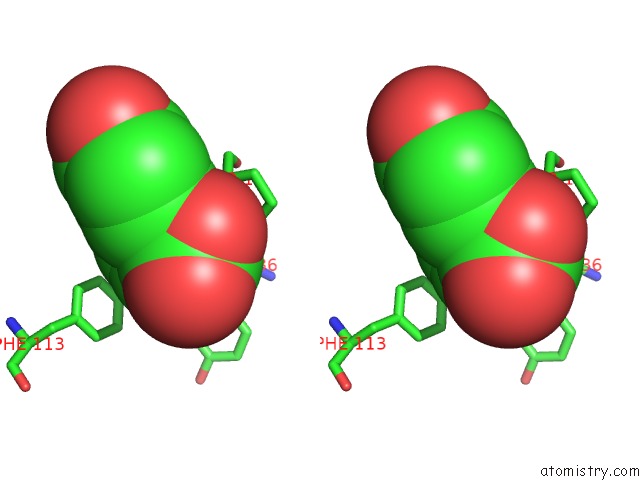
Fluorine binding site 1 out of 3 in 1mfi
Go back to
Fluorine binding site 1 out
of 3 in the Crystal Structure of Macrophage Migration Inhibitory Factor Complexed with (E)-2-Fluoro-P-Hydroxycinnamate

Mono view

Stereo pair view
Mono view
Stereo pair view
A full contact list of Fluorine with other atoms in the F binding
site number 1 of Crystal Structure of Macrophage Migration Inhibitory Factor Complexed with (E)-2-Fluoro-P-Hydroxycinnamate within 5.0Å range:
|
Fluorine binding site 2 out of 3 in 1mfi
Go back to
Fluorine binding site 2 out
of 3 in the Crystal Structure of Macrophage Migration Inhibitory Factor Complexed with (E)-2-Fluoro-P-Hydroxycinnamate

Mono view
Stereo pair view
Mono view
Stereo pair view
A full contact list of Fluorine with other atoms in the F binding
site number 2 of Crystal Structure of Macrophage Migration Inhibitory Factor Complexed with (E)-2-Fluoro-P-Hydroxycinnamate within 5.0Å range:
|
Fluorine binding site 3 out of 3 in 1mfi
Go back to
Fluorine binding site 3 out
of 3 in the Crystal Structure of Macrophage Migration Inhibitory Factor Complexed with (E)-2-Fluoro-P-Hydroxycinnamate

Mono view

Stereo pair view
Mono view
Stereo pair view
A full contact list of Fluorine with other atoms in the F binding
site number 3 of Crystal Structure of Macrophage Migration Inhibitory Factor Complexed with (E)-2-Fluoro-P-Hydroxycinnamate within 5.0Å range:
|
Reference:
A.B.Taylor,
W.H.Johnson Jr.,
R.M.Czerwinski,
H.S.Li,
M.L.Hackert,
C.P.Whitman.
Crystal Structure of Macrophage Migration Inhibitory Factor Complexed with (E)-2-Fluoro-P-Hydroxycinnamate at 1.8 A Resolution: Implications For Enzymatic Catalysis and Inhibition. Biochemistry V. 38 7444 1999.
ISSN: ISSN 0006-2960
PubMed: 10360941
DOI: 10.1021/BI9904048
Page generated: Wed Jul 31 11:51:40 2024
ISSN: ISSN 0006-2960
PubMed: 10360941
DOI: 10.1021/BI9904048
Last articles
Zn in 9J0NZn in 9J0O
Zn in 9J0P
Zn in 9FJX
Zn in 9EKB
Zn in 9C0F
Zn in 9CAH
Zn in 9CH0
Zn in 9CH3
Zn in 9CH1