Fluorine »
PDB 1o5g-1q6m »
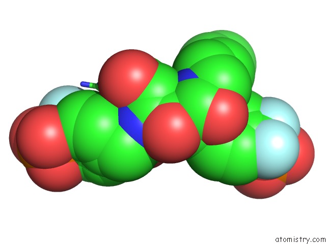
1pxh »
Fluorine in PDB 1pxh: Crystal Structure of Protein Tyrosine Phosphatase 1B with Potent and Selective Bidentate Inhibitor Compound 2
Enzymatic activity of Crystal Structure of Protein Tyrosine Phosphatase 1B with Potent and Selective Bidentate Inhibitor Compound 2
All present enzymatic activity of Crystal Structure of Protein Tyrosine Phosphatase 1B with Potent and Selective Bidentate Inhibitor Compound 2:
3.1.3.48;
3.1.3.48;
Protein crystallography data
The structure of Crystal Structure of Protein Tyrosine Phosphatase 1B with Potent and Selective Bidentate Inhibitor Compound 2, PDB code: 1pxh
was solved by
J.P.Sun,
A.Fedorov,
S.Y.Lee,
X.L.Guo,
K.Shen,
D.S.Lawrence,
S.C.Almo,
Z.Y.Zhang,
with X-Ray Crystallography technique. A brief refinement statistics is given in the table below:
| Resolution Low / High (Å) | 20.00 / 2.15 |
| Space group | P 21 21 21 |
| Cell size a, b, c (Å), α, β, γ (°) | 52.840, 85.678, 88.678, 90.00, 90.00, 90.00 |
| R / Rfree (%) | 20 / 20.7 |
Other elements in 1pxh:
The structure of Crystal Structure of Protein Tyrosine Phosphatase 1B with Potent and Selective Bidentate Inhibitor Compound 2 also contains other interesting chemical elements:
| Magnesium | (Mg) | 2 atoms |
Fluorine Binding Sites:
The binding sites of Fluorine atom in the Crystal Structure of Protein Tyrosine Phosphatase 1B with Potent and Selective Bidentate Inhibitor Compound 2
(pdb code 1pxh). This binding sites where shown within
5.0 Angstroms radius around Fluorine atom.
In total 4 binding sites of Fluorine where determined in the Crystal Structure of Protein Tyrosine Phosphatase 1B with Potent and Selective Bidentate Inhibitor Compound 2, PDB code: 1pxh:
Jump to Fluorine binding site number: 1; 2; 3; 4;
In total 4 binding sites of Fluorine where determined in the Crystal Structure of Protein Tyrosine Phosphatase 1B with Potent and Selective Bidentate Inhibitor Compound 2, PDB code: 1pxh:
Jump to Fluorine binding site number: 1; 2; 3; 4;
Fluorine binding site 1 out of 4 in 1pxh
Go back to
Fluorine binding site 1 out
of 4 in the Crystal Structure of Protein Tyrosine Phosphatase 1B with Potent and Selective Bidentate Inhibitor Compound 2

Mono view
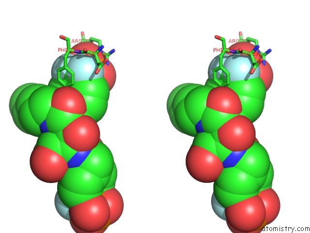
Stereo pair view
Mono view
Stereo pair view
A full contact list of Fluorine with other atoms in the F binding
site number 1 of Crystal Structure of Protein Tyrosine Phosphatase 1B with Potent and Selective Bidentate Inhibitor Compound 2 within 5.0Å range:
|
Fluorine binding site 2 out of 4 in 1pxh
Go back to
Fluorine binding site 2 out
of 4 in the Crystal Structure of Protein Tyrosine Phosphatase 1B with Potent and Selective Bidentate Inhibitor Compound 2
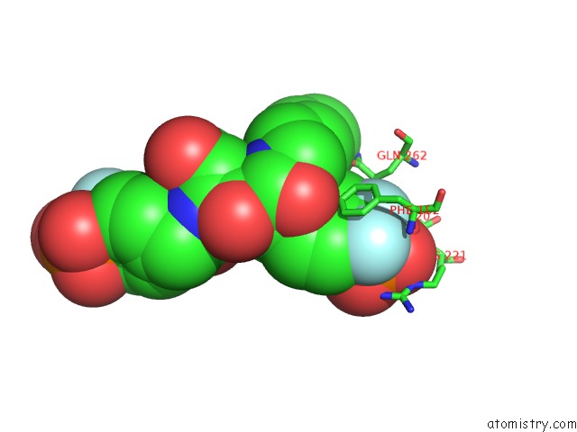
Mono view
Stereo pair view
Mono view
Stereo pair view
A full contact list of Fluorine with other atoms in the F binding
site number 2 of Crystal Structure of Protein Tyrosine Phosphatase 1B with Potent and Selective Bidentate Inhibitor Compound 2 within 5.0Å range:
|
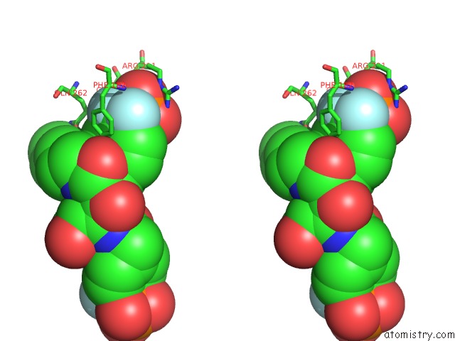
Fluorine binding site 3 out of 4 in 1pxh
Go back to
Fluorine binding site 3 out
of 4 in the Crystal Structure of Protein Tyrosine Phosphatase 1B with Potent and Selective Bidentate Inhibitor Compound 2
Mono view

Stereo pair view
Mono view
Stereo pair view
A full contact list of Fluorine with other atoms in the F binding
site number 3 of Crystal Structure of Protein Tyrosine Phosphatase 1B with Potent and Selective Bidentate Inhibitor Compound 2 within 5.0Å range:
|
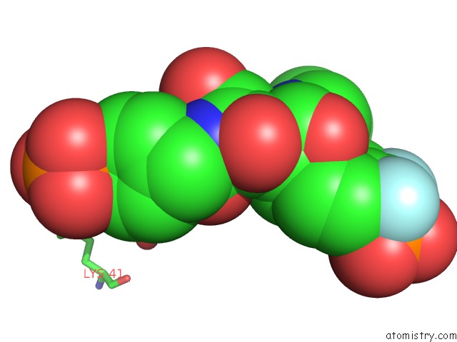
Fluorine binding site 4 out of 4 in 1pxh
Go back to
Fluorine binding site 4 out
of 4 in the Crystal Structure of Protein Tyrosine Phosphatase 1B with Potent and Selective Bidentate Inhibitor Compound 2
Mono view

Stereo pair view
Mono view
Stereo pair view
A full contact list of Fluorine with other atoms in the F binding
site number 4 of Crystal Structure of Protein Tyrosine Phosphatase 1B with Potent and Selective Bidentate Inhibitor Compound 2 within 5.0Å range:
|
Reference:
J.P.Sun,
A.A.Fedorov,
S.Y.Lee,
X.L.Guo,
K.Shen,
D.S.Lawrence,
S.C.Almo,
Z.Y.Zhang.
Crystal Structure of PTP1B Complexed with A Potent and Selective Bidentate Inhibitor. J.Biol.Chem. V. 278 12406 2003.
ISSN: ISSN 0021-9258
PubMed: 12547827
DOI: 10.1074/JBC.M212491200
Page generated: Wed Jul 31 12:22:27 2024
ISSN: ISSN 0021-9258
PubMed: 12547827
DOI: 10.1074/JBC.M212491200
Last articles
Zn in 9JYWZn in 9IR4
Zn in 9IR3
Zn in 9GMX
Zn in 9GMW
Zn in 9JEJ
Zn in 9ERF
Zn in 9ERE
Zn in 9EGV
Zn in 9EGW