Fluorine »
PDB 1q6n-1rrx »
1q6p »
Fluorine in PDB 1q6p: The Structure of Phosphotyrosine Phosphatase 1B in Complex with Compound 6
Enzymatic activity of The Structure of Phosphotyrosine Phosphatase 1B in Complex with Compound 6
All present enzymatic activity of The Structure of Phosphotyrosine Phosphatase 1B in Complex with Compound 6:
3.1.3.48;
3.1.3.48;
Protein crystallography data
The structure of The Structure of Phosphotyrosine Phosphatase 1B in Complex with Compound 6, PDB code: 1q6p
was solved by
G.Scapin,
S.B.Patel,
J.W.Becker,
Q.Wang,
C.Desponts,
D.Waddleton,
K.Skorey,
W.Cromlish,
C.Bayly,
M.Therien,
J.Y.Gauthier,
C.S.Li,
C.K.Lau,
C.Ramachandran,
B.P.Kennedy,
E.Asante-Appiah,
with X-Ray Crystallography technique. A brief refinement statistics is given in the table below:
| Resolution Low / High (Å) | 15.00 / 2.30 |
| Space group | P 21 21 21 |
| Cell size a, b, c (Å), α, β, γ (°) | 87.945, 88.045, 139.228, 90.00, 90.00, 90.00 |
| R / Rfree (%) | n/a / n/a |
Other elements in 1q6p:
The structure of The Structure of Phosphotyrosine Phosphatase 1B in Complex with Compound 6 also contains other interesting chemical elements:
| Chlorine | (Cl) | 1 atom |
Fluorine Binding Sites:
The binding sites of Fluorine atom in the The Structure of Phosphotyrosine Phosphatase 1B in Complex with Compound 6
(pdb code 1q6p). This binding sites where shown within
5.0 Angstroms radius around Fluorine atom.
In total 4 binding sites of Fluorine where determined in the The Structure of Phosphotyrosine Phosphatase 1B in Complex with Compound 6, PDB code: 1q6p:
Jump to Fluorine binding site number: 1; 2; 3; 4;
In total 4 binding sites of Fluorine where determined in the The Structure of Phosphotyrosine Phosphatase 1B in Complex with Compound 6, PDB code: 1q6p:
Jump to Fluorine binding site number: 1; 2; 3; 4;
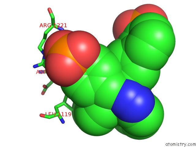
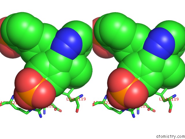
Fluorine binding site 1 out of 4 in 1q6p
Go back to
Fluorine binding site 1 out
of 4 in the The Structure of Phosphotyrosine Phosphatase 1B in Complex with Compound 6
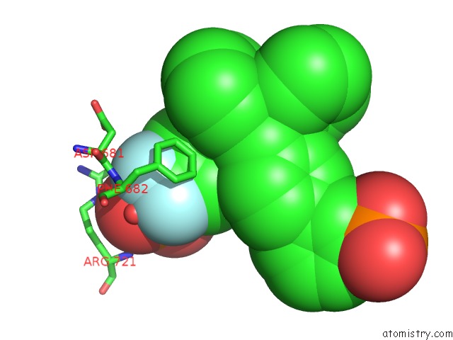
Mono view
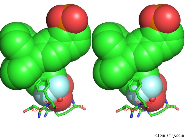
Stereo pair view
Mono view
Stereo pair view
A full contact list of Fluorine with other atoms in the F binding
site number 1 of The Structure of Phosphotyrosine Phosphatase 1B in Complex with Compound 6 within 5.0Å range:
|
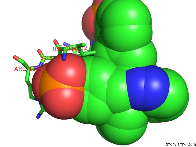
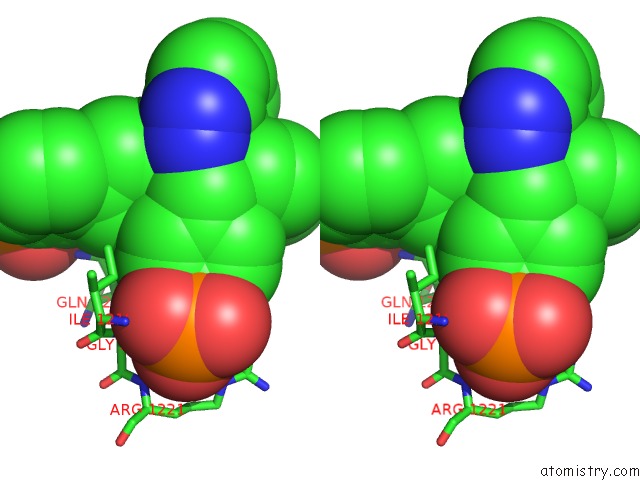
Fluorine binding site 2 out of 4 in 1q6p
Go back to
Fluorine binding site 2 out
of 4 in the The Structure of Phosphotyrosine Phosphatase 1B in Complex with Compound 6
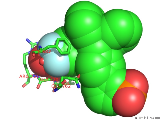
Mono view
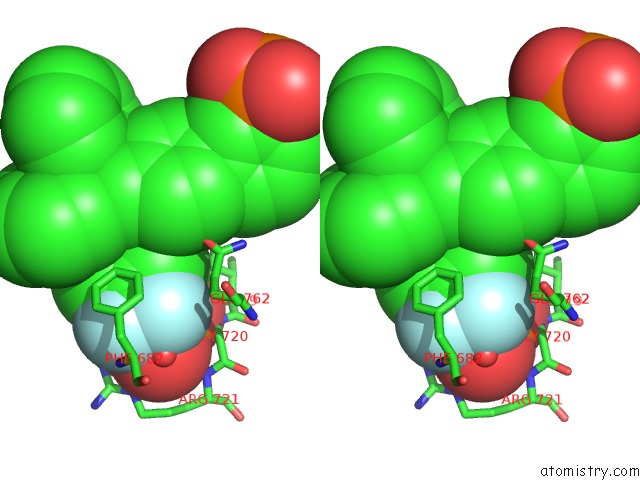
Stereo pair view
Mono view
Stereo pair view
A full contact list of Fluorine with other atoms in the F binding
site number 2 of The Structure of Phosphotyrosine Phosphatase 1B in Complex with Compound 6 within 5.0Å range:
|
Fluorine binding site 3 out of 4 in 1q6p
Go back to
Fluorine binding site 3 out
of 4 in the The Structure of Phosphotyrosine Phosphatase 1B in Complex with Compound 6
Mono view
Stereo pair view
Mono view
Stereo pair view
A full contact list of Fluorine with other atoms in the F binding
site number 3 of The Structure of Phosphotyrosine Phosphatase 1B in Complex with Compound 6 within 5.0Å range:
|
Fluorine binding site 4 out of 4 in 1q6p
Go back to
Fluorine binding site 4 out
of 4 in the The Structure of Phosphotyrosine Phosphatase 1B in Complex with Compound 6
Mono view
Stereo pair view
Mono view
Stereo pair view
A full contact list of Fluorine with other atoms in the F binding
site number 4 of The Structure of Phosphotyrosine Phosphatase 1B in Complex with Compound 6 within 5.0Å range:
|
Reference:
G.Scapin,
S.B.Patel,
J.W.Becker,
Q.Wang,
C.Desponts,
D.Waddleton,
K.Skorey,
W.Cromlish,
C.Bayly,
M.Therien,
J.Y.Gauthier,
C.S.Li,
C.K.Lau,
C.Ramachandran,
B.P.Kennedy,
E.Asante-Appiah.
The Structural Basis For the Selectivity of Benzotriazole Inhibitors of PTP1B Biochemistry V. 42 11451 2003.
ISSN: ISSN 0006-2960
PubMed: 14516196
DOI: 10.1021/BI035098J
Page generated: Wed Jul 31 12:32:11 2024
ISSN: ISSN 0006-2960
PubMed: 14516196
DOI: 10.1021/BI035098J
Last articles
Zn in 9J0NZn in 9J0O
Zn in 9J0P
Zn in 9FJX
Zn in 9EKB
Zn in 9C0F
Zn in 9CAH
Zn in 9CH0
Zn in 9CH3
Zn in 9CH1