Fluorine »
PDB 1rw8-1uda »
1ta0 »
Fluorine in PDB 1ta0: Three-Dimensional Structure of A Rna-Polymerase II Binding Protein with Associated Ligand.
Enzymatic activity of Three-Dimensional Structure of A Rna-Polymerase II Binding Protein with Associated Ligand.
All present enzymatic activity of Three-Dimensional Structure of A Rna-Polymerase II Binding Protein with Associated Ligand.:
3.1.3.16;
3.1.3.16;
Protein crystallography data
The structure of Three-Dimensional Structure of A Rna-Polymerase II Binding Protein with Associated Ligand., PDB code: 1ta0
was solved by
T.Kamenski,
S.Heilmeier,
T.Meinhart,
P.Cramer,
with X-Ray Crystallography technique. A brief refinement statistics is given in the table below:
| Resolution Low / High (Å) | 20.00 / 2.10 |
| Space group | P 21 21 2 |
| Cell size a, b, c (Å), α, β, γ (°) | 117.820, 47.170, 40.070, 90.00, 90.00, 90.00 |
| R / Rfree (%) | 20.7 / 22.7 |
Other elements in 1ta0:
The structure of Three-Dimensional Structure of A Rna-Polymerase II Binding Protein with Associated Ligand. also contains other interesting chemical elements:
| Magnesium | (Mg) | 1 atom |
Fluorine Binding Sites:
The binding sites of Fluorine atom in the Three-Dimensional Structure of A Rna-Polymerase II Binding Protein with Associated Ligand.
(pdb code 1ta0). This binding sites where shown within
5.0 Angstroms radius around Fluorine atom.
In total 3 binding sites of Fluorine where determined in the Three-Dimensional Structure of A Rna-Polymerase II Binding Protein with Associated Ligand., PDB code: 1ta0:
Jump to Fluorine binding site number: 1; 2; 3;
In total 3 binding sites of Fluorine where determined in the Three-Dimensional Structure of A Rna-Polymerase II Binding Protein with Associated Ligand., PDB code: 1ta0:
Jump to Fluorine binding site number: 1; 2; 3;
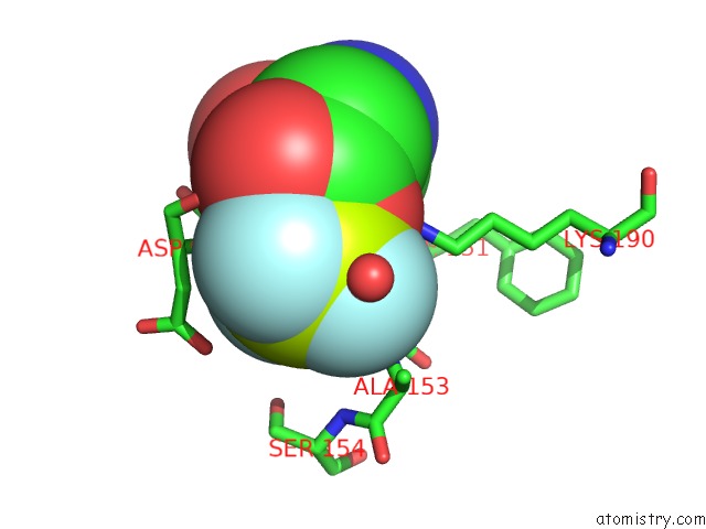
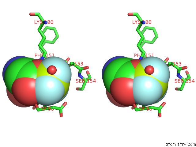
Fluorine binding site 1 out of 3 in 1ta0
Go back to
Fluorine binding site 1 out
of 3 in the Three-Dimensional Structure of A Rna-Polymerase II Binding Protein with Associated Ligand.
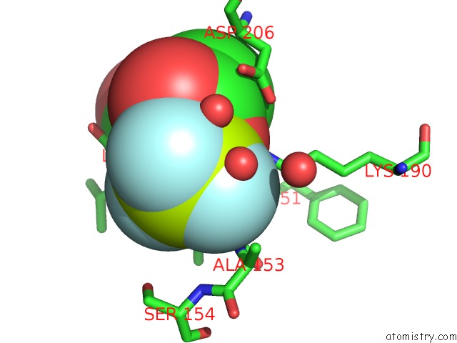
Mono view
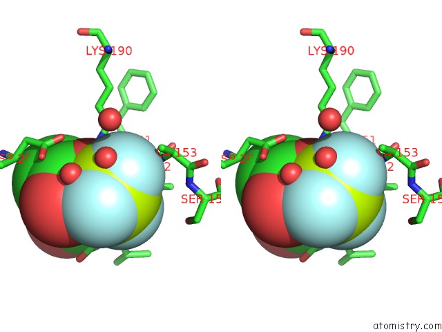
Stereo pair view
Mono view
Stereo pair view
A full contact list of Fluorine with other atoms in the F binding
site number 1 of Three-Dimensional Structure of A Rna-Polymerase II Binding Protein with Associated Ligand. within 5.0Å range:
|
Fluorine binding site 2 out of 3 in 1ta0
Go back to
Fluorine binding site 2 out
of 3 in the Three-Dimensional Structure of A Rna-Polymerase II Binding Protein with Associated Ligand.
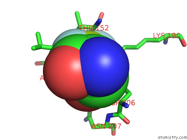
Mono view
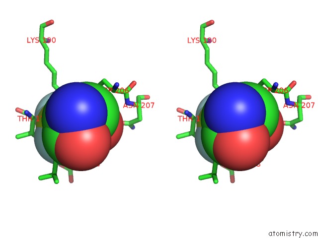
Stereo pair view
Mono view
Stereo pair view
A full contact list of Fluorine with other atoms in the F binding
site number 2 of Three-Dimensional Structure of A Rna-Polymerase II Binding Protein with Associated Ligand. within 5.0Å range:
|
Fluorine binding site 3 out of 3 in 1ta0
Go back to
Fluorine binding site 3 out
of 3 in the Three-Dimensional Structure of A Rna-Polymerase II Binding Protein with Associated Ligand.
Mono view
Stereo pair view
Mono view
Stereo pair view
A full contact list of Fluorine with other atoms in the F binding
site number 3 of Three-Dimensional Structure of A Rna-Polymerase II Binding Protein with Associated Ligand. within 5.0Å range:
|
Reference:
T.Kamenski,
S.Heilmeier,
T.Meinhart,
P.Cramer.
Structure and Mechanism of Rna Polymerase II Ctd Phosphatases. Mol.Cell V. 15 399 2004.
ISSN: ISSN 1097-2765
PubMed: 15304220
DOI: 10.1016/J.MOLCEL.2004.06.035
Page generated: Wed Jul 31 12:56:35 2024
ISSN: ISSN 1097-2765
PubMed: 15304220
DOI: 10.1016/J.MOLCEL.2004.06.035
Last articles
Zn in 9J0NZn in 9J0O
Zn in 9J0P
Zn in 9FJX
Zn in 9EKB
Zn in 9C0F
Zn in 9CAH
Zn in 9CH0
Zn in 9CH3
Zn in 9CH1