Fluorine »
PDB 1udb-1w5y »
1uhv »
Fluorine in PDB 1uhv: Crystal Structure of Beta-D-Xylosidase From Thermoanaerobacterium Saccharolyticum, A Family 39 Glycoside Hydrolase
Enzymatic activity of Crystal Structure of Beta-D-Xylosidase From Thermoanaerobacterium Saccharolyticum, A Family 39 Glycoside Hydrolase
All present enzymatic activity of Crystal Structure of Beta-D-Xylosidase From Thermoanaerobacterium Saccharolyticum, A Family 39 Glycoside Hydrolase:
3.2.1.37;
3.2.1.37;
Protein crystallography data
The structure of Crystal Structure of Beta-D-Xylosidase From Thermoanaerobacterium Saccharolyticum, A Family 39 Glycoside Hydrolase, PDB code: 1uhv
was solved by
J.K.Yang,
H.J.Yoon,
H.J.Ahn,
B.Il Lee,
J.D.Pedelacq,
E.C.Liong,
J.Berendzen,
M.Laivenieks,
C.Vieille,
G.J.Zeikus,
D.J.Vocadlo,
S.G.Withers,
S.W.Suh,
with X-Ray Crystallography technique. A brief refinement statistics is given in the table below:
| Resolution Low / High (Å) | 32.33 / 2.10 |
| Space group | P 21 21 21 |
| Cell size a, b, c (Å), α, β, γ (°) | 95.326, 152.276, 159.684, 90.00, 90.00, 90.00 |
| R / Rfree (%) | 21 / 26.2 |
Fluorine Binding Sites:
The binding sites of Fluorine atom in the Crystal Structure of Beta-D-Xylosidase From Thermoanaerobacterium Saccharolyticum, A Family 39 Glycoside Hydrolase
(pdb code 1uhv). This binding sites where shown within
5.0 Angstroms radius around Fluorine atom.
In total 4 binding sites of Fluorine where determined in the Crystal Structure of Beta-D-Xylosidase From Thermoanaerobacterium Saccharolyticum, A Family 39 Glycoside Hydrolase, PDB code: 1uhv:
Jump to Fluorine binding site number: 1; 2; 3; 4;
In total 4 binding sites of Fluorine where determined in the Crystal Structure of Beta-D-Xylosidase From Thermoanaerobacterium Saccharolyticum, A Family 39 Glycoside Hydrolase, PDB code: 1uhv:
Jump to Fluorine binding site number: 1; 2; 3; 4;
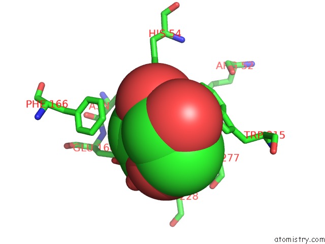
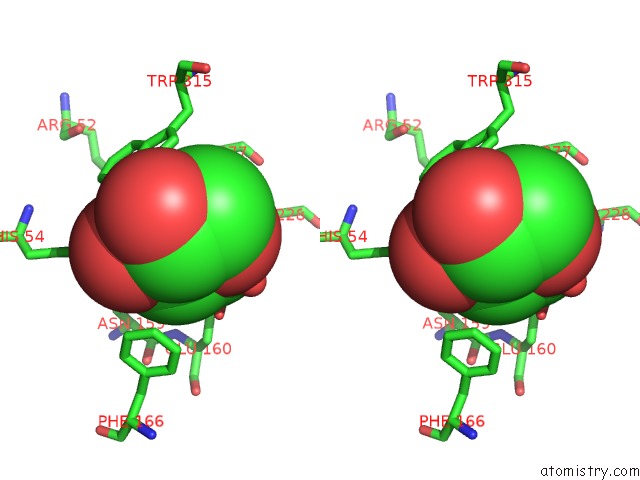
Fluorine binding site 1 out of 4 in 1uhv
Go back to
Fluorine binding site 1 out
of 4 in the Crystal Structure of Beta-D-Xylosidase From Thermoanaerobacterium Saccharolyticum, A Family 39 Glycoside Hydrolase
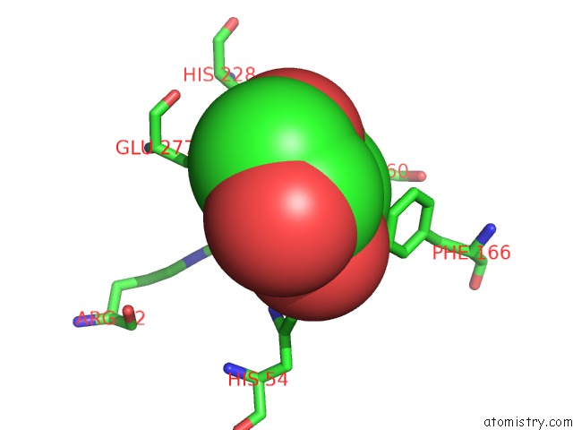
Mono view
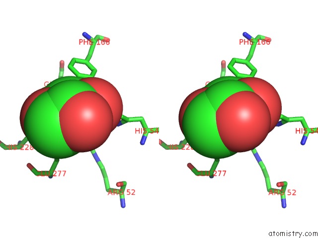
Stereo pair view
Mono view
Stereo pair view
A full contact list of Fluorine with other atoms in the F binding
site number 1 of Crystal Structure of Beta-D-Xylosidase From Thermoanaerobacterium Saccharolyticum, A Family 39 Glycoside Hydrolase within 5.0Å range:
|
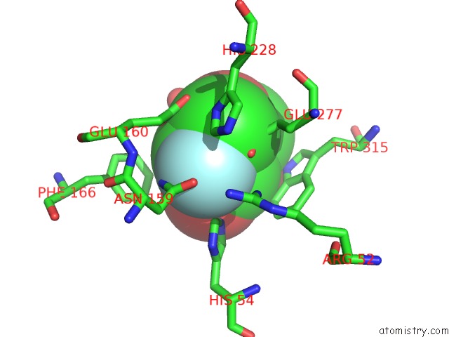
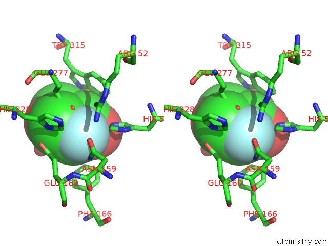
Fluorine binding site 2 out of 4 in 1uhv
Go back to
Fluorine binding site 2 out
of 4 in the Crystal Structure of Beta-D-Xylosidase From Thermoanaerobacterium Saccharolyticum, A Family 39 Glycoside Hydrolase
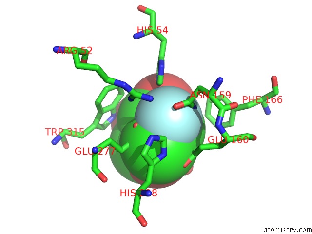
Mono view
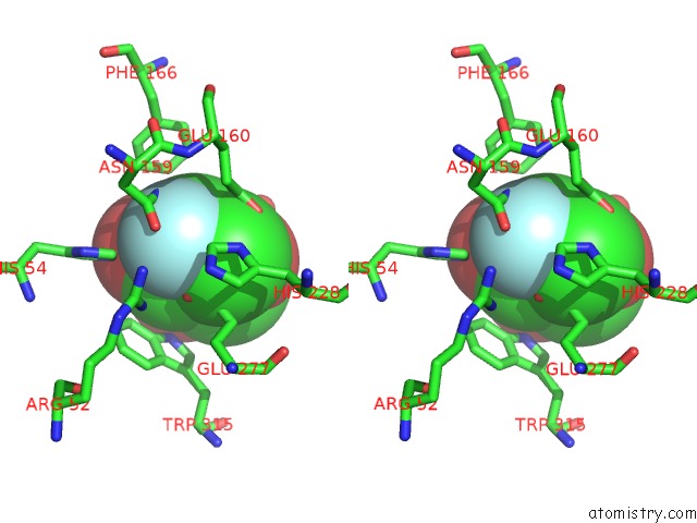
Stereo pair view
Mono view
Stereo pair view
A full contact list of Fluorine with other atoms in the F binding
site number 2 of Crystal Structure of Beta-D-Xylosidase From Thermoanaerobacterium Saccharolyticum, A Family 39 Glycoside Hydrolase within 5.0Å range:
|
Fluorine binding site 3 out of 4 in 1uhv
Go back to
Fluorine binding site 3 out
of 4 in the Crystal Structure of Beta-D-Xylosidase From Thermoanaerobacterium Saccharolyticum, A Family 39 Glycoside Hydrolase
Mono view
Stereo pair view
Mono view
Stereo pair view
A full contact list of Fluorine with other atoms in the F binding
site number 3 of Crystal Structure of Beta-D-Xylosidase From Thermoanaerobacterium Saccharolyticum, A Family 39 Glycoside Hydrolase within 5.0Å range:
|
Fluorine binding site 4 out of 4 in 1uhv
Go back to
Fluorine binding site 4 out
of 4 in the Crystal Structure of Beta-D-Xylosidase From Thermoanaerobacterium Saccharolyticum, A Family 39 Glycoside Hydrolase
Mono view
Stereo pair view
Mono view
Stereo pair view
A full contact list of Fluorine with other atoms in the F binding
site number 4 of Crystal Structure of Beta-D-Xylosidase From Thermoanaerobacterium Saccharolyticum, A Family 39 Glycoside Hydrolase within 5.0Å range:
|
Reference:
J.K.Yang,
H.J.Yoon,
H.J.Ahn,
B.I.Lee,
J.D.Pedelacq,
E.C.Liong,
J.Berendzen,
M.Laivenieks,
C.Vieille,
G.J.Zeikus,
D.J.Vocadlo,
S.G.Withers,
S.W.Suh.
Crystal Structure of Beta-D-Xylosidase From Thermoanaerobacterium Saccharolyticum, A Family 39 Glycoside Hydrolase. J.Mol.Biol. V. 335 155 2004.
ISSN: ISSN 0022-2836
PubMed: 14659747
DOI: 10.1016/J.JMB.2003.10.026
Page generated: Wed Jul 31 13:02:22 2024
ISSN: ISSN 0022-2836
PubMed: 14659747
DOI: 10.1016/J.JMB.2003.10.026
Last articles
Zn in 9J0NZn in 9J0O
Zn in 9J0P
Zn in 9FJX
Zn in 9EKB
Zn in 9C0F
Zn in 9CAH
Zn in 9CH0
Zn in 9CH3
Zn in 9CH1