Fluorine »
PDB 1udb-1w5y »
1veb »
Fluorine in PDB 1veb: Crystal Structure of Protein Kinase A in Complex with Azepane Derivative 5
Enzymatic activity of Crystal Structure of Protein Kinase A in Complex with Azepane Derivative 5
All present enzymatic activity of Crystal Structure of Protein Kinase A in Complex with Azepane Derivative 5:
2.7.1.37;
2.7.1.37;
Protein crystallography data
The structure of Crystal Structure of Protein Kinase A in Complex with Azepane Derivative 5, PDB code: 1veb
was solved by
C.B.Breitenlechner,
T.Wegge,
L.Berillon,
K.Graul,
K.Marzenell,
W.-G.Friebe,
U.Thomas,
R.Schumacher,
R.Huber,
R.A.Engh,
B.Masjost,
with X-Ray Crystallography technique. A brief refinement statistics is given in the table below:
| Resolution Low / High (Å) | 21.52 / 2.89 |
| Space group | P 21 21 21 |
| Cell size a, b, c (Å), α, β, γ (°) | 74.881, 76.510, 80.094, 90.00, 90.00, 90.00 |
| R / Rfree (%) | 18.3 / 25.1 |
Fluorine Binding Sites:
The binding sites of Fluorine atom in the Crystal Structure of Protein Kinase A in Complex with Azepane Derivative 5
(pdb code 1veb). This binding sites where shown within
5.0 Angstroms radius around Fluorine atom.
In total only one binding site of Fluorine was determined in the Crystal Structure of Protein Kinase A in Complex with Azepane Derivative 5, PDB code: 1veb:
In total only one binding site of Fluorine was determined in the Crystal Structure of Protein Kinase A in Complex with Azepane Derivative 5, PDB code: 1veb:
Fluorine binding site 1 out of 1 in 1veb
Go back to
Fluorine binding site 1 out
of 1 in the Crystal Structure of Protein Kinase A in Complex with Azepane Derivative 5
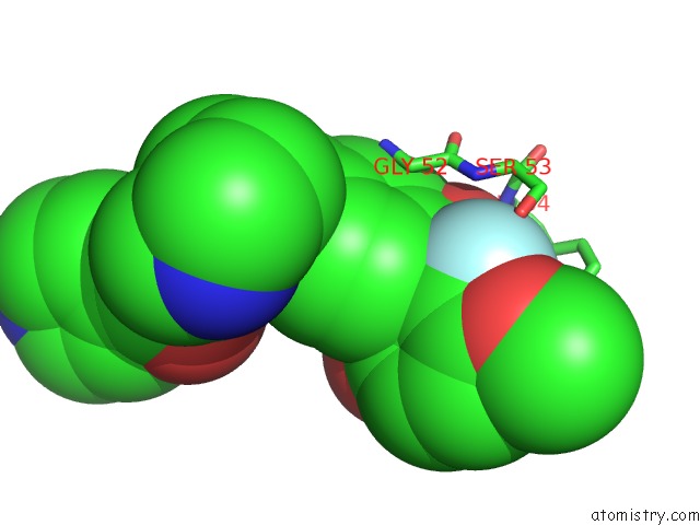
Mono view
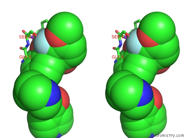
Stereo pair view
Mono view
Stereo pair view
A full contact list of Fluorine with other atoms in the F binding
site number 1 of Crystal Structure of Protein Kinase A in Complex with Azepane Derivative 5 within 5.0Å range:
|
Reference:
C.B.Breitenlechner,
T.Wegge,
L.Berillon,
K.Graul,
K.Marzenell,
W.-G.Friebe,
U.Thomas,
R.Schumacher,
R.Huber,
R.A.Engh,
B.Masjost.
Structure-Based Optimization of Novel Azepane Derivatives As Pkb Inhibitors J.Med.Chem. V. 47 1375 2004.
ISSN: ISSN 0022-2623
PubMed: 14998327
DOI: 10.1021/JM0310479
Page generated: Wed Jul 31 13:08:31 2024
ISSN: ISSN 0022-2623
PubMed: 14998327
DOI: 10.1021/JM0310479
Last articles
Zn in 9J0NZn in 9J0O
Zn in 9J0P
Zn in 9FJX
Zn in 9EKB
Zn in 9C0F
Zn in 9CAH
Zn in 9CH0
Zn in 9CH3
Zn in 9CH1