Fluorine »
PDB 1w6j-1xz1 »
1w9i »
Fluorine in PDB 1w9i: Myosin II Dictyostelium Discoideum Motor Domain S456Y Bound with Mgadp-Befx
Enzymatic activity of Myosin II Dictyostelium Discoideum Motor Domain S456Y Bound with Mgadp-Befx
All present enzymatic activity of Myosin II Dictyostelium Discoideum Motor Domain S456Y Bound with Mgadp-Befx:
3.6.4.1;
3.6.4.1;
Protein crystallography data
The structure of Myosin II Dictyostelium Discoideum Motor Domain S456Y Bound with Mgadp-Befx, PDB code: 1w9i
was solved by
C.A.Morris,
P.-D.Coureux,
A.L.Wells,
A.Houdusse,
H.L.Sweeney,
with X-Ray Crystallography technique. A brief refinement statistics is given in the table below:
| Resolution Low / High (Å) | 40.00 / 1.75 |
| Space group | P 21 21 2 |
| Cell size a, b, c (Å), α, β, γ (°) | 105.017, 186.506, 54.713, 90.00, 90.00, 90.00 |
| R / Rfree (%) | 18.2 / 20.7 |
Other elements in 1w9i:
The structure of Myosin II Dictyostelium Discoideum Motor Domain S456Y Bound with Mgadp-Befx also contains other interesting chemical elements:
| Magnesium | (Mg) | 1 atom |
Fluorine Binding Sites:
The binding sites of Fluorine atom in the Myosin II Dictyostelium Discoideum Motor Domain S456Y Bound with Mgadp-Befx
(pdb code 1w9i). This binding sites where shown within
5.0 Angstroms radius around Fluorine atom.
In total 3 binding sites of Fluorine where determined in the Myosin II Dictyostelium Discoideum Motor Domain S456Y Bound with Mgadp-Befx, PDB code: 1w9i:
Jump to Fluorine binding site number: 1; 2; 3;
In total 3 binding sites of Fluorine where determined in the Myosin II Dictyostelium Discoideum Motor Domain S456Y Bound with Mgadp-Befx, PDB code: 1w9i:
Jump to Fluorine binding site number: 1; 2; 3;
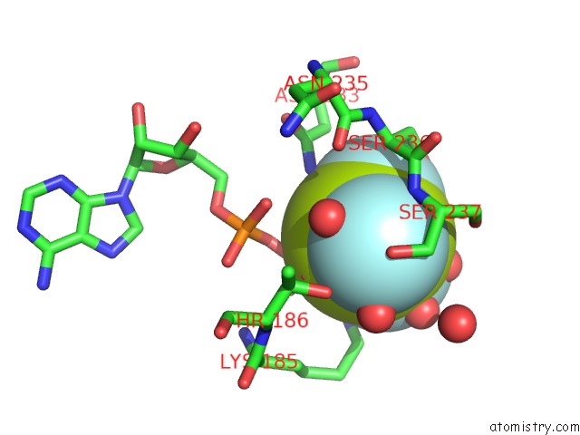
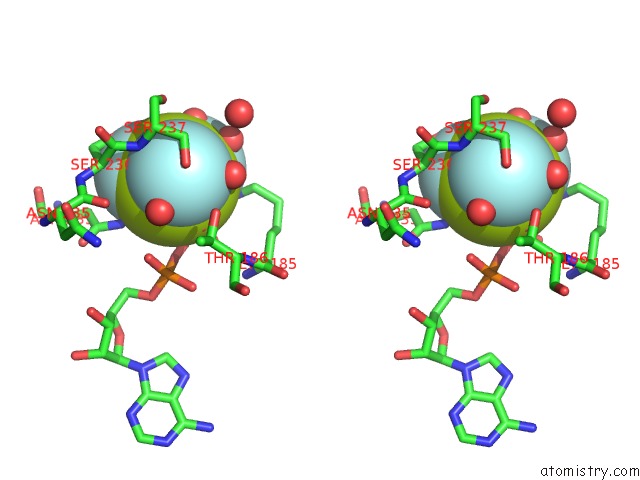
Fluorine binding site 1 out of 3 in 1w9i
Go back to
Fluorine binding site 1 out
of 3 in the Myosin II Dictyostelium Discoideum Motor Domain S456Y Bound with Mgadp-Befx
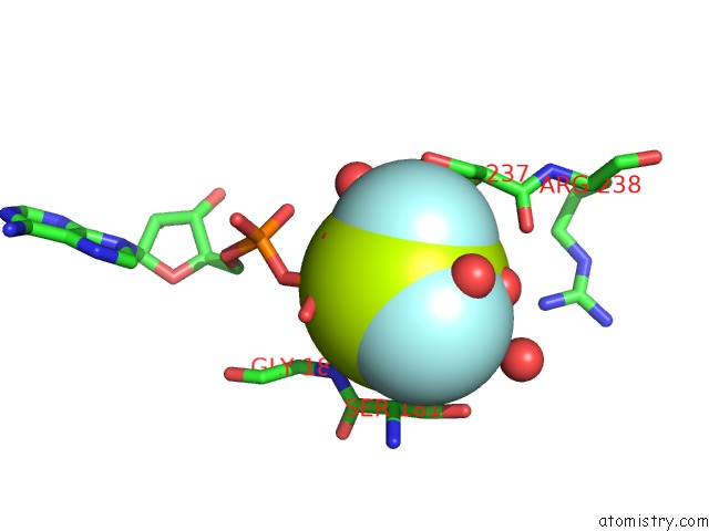
Mono view
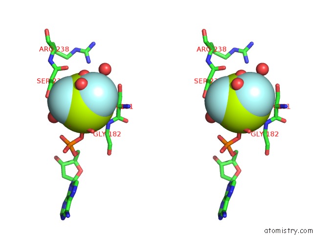
Stereo pair view
Mono view
Stereo pair view
A full contact list of Fluorine with other atoms in the F binding
site number 1 of Myosin II Dictyostelium Discoideum Motor Domain S456Y Bound with Mgadp-Befx within 5.0Å range:
|
Fluorine binding site 2 out of 3 in 1w9i
Go back to
Fluorine binding site 2 out
of 3 in the Myosin II Dictyostelium Discoideum Motor Domain S456Y Bound with Mgadp-Befx
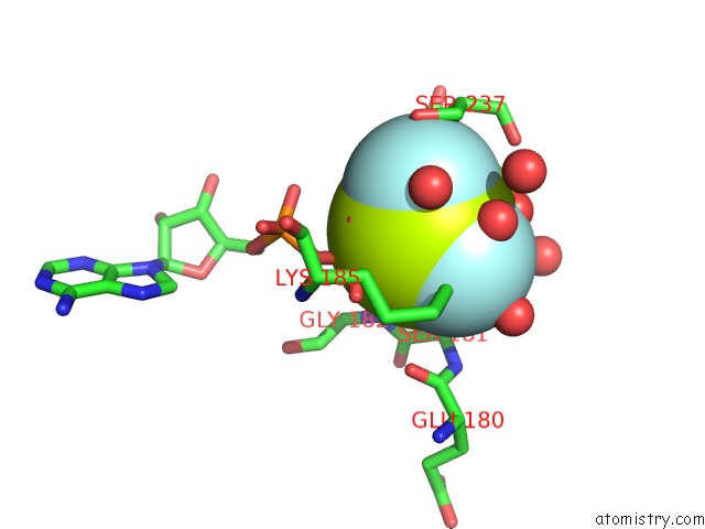
Mono view
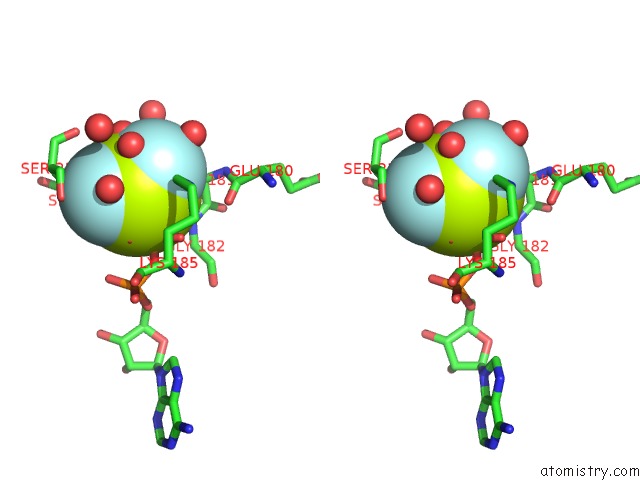
Stereo pair view
Mono view
Stereo pair view
A full contact list of Fluorine with other atoms in the F binding
site number 2 of Myosin II Dictyostelium Discoideum Motor Domain S456Y Bound with Mgadp-Befx within 5.0Å range:
|
Fluorine binding site 3 out of 3 in 1w9i
Go back to
Fluorine binding site 3 out
of 3 in the Myosin II Dictyostelium Discoideum Motor Domain S456Y Bound with Mgadp-Befx
Mono view
Stereo pair view
Mono view
Stereo pair view
A full contact list of Fluorine with other atoms in the F binding
site number 3 of Myosin II Dictyostelium Discoideum Motor Domain S456Y Bound with Mgadp-Befx within 5.0Å range:
|
Reference:
C.A.Morris,
P.-D.Coureux,
A.L.Wells,
A.Houdusse,
H.L.Sweeney.
Structure-Function Analysis of Myosin II Backdoor Mutants To Be Published.
Page generated: Wed Jul 31 13:13:38 2024
Last articles
Zn in 9J0NZn in 9J0O
Zn in 9J0P
Zn in 9FJX
Zn in 9EKB
Zn in 9C0F
Zn in 9CAH
Zn in 9CH0
Zn in 9CH3
Zn in 9CH1