Fluorine »
PDB 1w6j-1xz1 »
1xha »
Fluorine in PDB 1xha: Crystal Structures of Protein Kinase B Selective Inhibitors in Complex with Protein Kinase A and Mutants
Enzymatic activity of Crystal Structures of Protein Kinase B Selective Inhibitors in Complex with Protein Kinase A and Mutants
All present enzymatic activity of Crystal Structures of Protein Kinase B Selective Inhibitors in Complex with Protein Kinase A and Mutants:
2.7.1.37;
2.7.1.37;
Protein crystallography data
The structure of Crystal Structures of Protein Kinase B Selective Inhibitors in Complex with Protein Kinase A and Mutants, PDB code: 1xha
was solved by
C.B.Breitenlechner,
W.-G.Friebe,
E.Brunet,
G.Werner,
K.Graul,
U.Thomas,
K.-P.Kuenkele,
W.Schaefer,
M.Gassel,
D.Bossemeyer,
R.Huber,
R.A.Engh,
B.Masjost,
with X-Ray Crystallography technique. A brief refinement statistics is given in the table below:
| Resolution Low / High (Å) | 15.00 / 2.46 |
| Space group | P 21 21 21 |
| Cell size a, b, c (Å), α, β, γ (°) | 49.842, 79.572, 117.329, 90.00, 90.00, 90.00 |
| R / Rfree (%) | 22 / 27.2 |
Fluorine Binding Sites:
The binding sites of Fluorine atom in the Crystal Structures of Protein Kinase B Selective Inhibitors in Complex with Protein Kinase A and Mutants
(pdb code 1xha). This binding sites where shown within
5.0 Angstroms radius around Fluorine atom.
In total only one binding site of Fluorine was determined in the Crystal Structures of Protein Kinase B Selective Inhibitors in Complex with Protein Kinase A and Mutants, PDB code: 1xha:
In total only one binding site of Fluorine was determined in the Crystal Structures of Protein Kinase B Selective Inhibitors in Complex with Protein Kinase A and Mutants, PDB code: 1xha:
Fluorine binding site 1 out of 1 in 1xha
Go back to
Fluorine binding site 1 out
of 1 in the Crystal Structures of Protein Kinase B Selective Inhibitors in Complex with Protein Kinase A and Mutants

Mono view
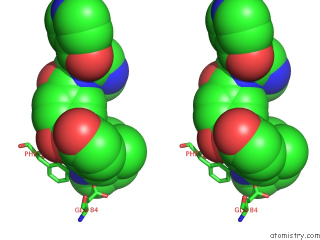
Stereo pair view
Mono view
Stereo pair view
A full contact list of Fluorine with other atoms in the F binding
site number 1 of Crystal Structures of Protein Kinase B Selective Inhibitors in Complex with Protein Kinase A and Mutants within 5.0Å range:
|
Reference:
C.B.Breitenlechner,
W.-G.Friebe,
E.Brunet,
G.Werner,
K.Graul,
U.Thomas,
K.-P.Kuenkele,
W.Schaefer,
M.Gassel,
D.Bossemeyer,
R.Huber,
R.A.Engh,
B.Masjost.
Design and Crystal Structures of Protein Kinase B-Selective Inhibitors in Complex with Protein Kinase A and Mutants J.Med.Chem. V. 48 163 2005.
ISSN: ISSN 0022-2623
PubMed: 15634010
DOI: 10.1021/JM049701N
Page generated: Wed Jul 31 13:18:14 2024
ISSN: ISSN 0022-2623
PubMed: 15634010
DOI: 10.1021/JM049701N
Last articles
Ca in 5TI8Ca in 5THG
Ca in 5TGX
Ca in 5TFK
Ca in 5TG3
Ca in 5TGQ
Ca in 5TGF
Ca in 5TFM
Ca in 5TEA
Ca in 5TFL