Fluorine »
PDB 1xz3-1zzr »
1zl3 »
Fluorine in PDB 1zl3: Coupling of Active Site Motions and Rna Binding
Enzymatic activity of Coupling of Active Site Motions and Rna Binding
All present enzymatic activity of Coupling of Active Site Motions and Rna Binding:
4.2.1.70;
4.2.1.70;
Protein crystallography data
The structure of Coupling of Active Site Motions and Rna Binding, PDB code: 1zl3
was solved by
C.Hoang,
C.S.Hamilton,
E.G.Mueller,
A.R.Ferre-D'amare,
with X-Ray Crystallography technique. A brief refinement statistics is given in the table below:
| Resolution Low / High (Å) | 19.92 / 2.80 |
| Space group | C 1 2 1 |
| Cell size a, b, c (Å), α, β, γ (°) | 145.433, 40.753, 78.803, 90.00, 110.61, 90.00 |
| R / Rfree (%) | 20.2 / 26.4 |
Fluorine Binding Sites:
The binding sites of Fluorine atom in the Coupling of Active Site Motions and Rna Binding
(pdb code 1zl3). This binding sites where shown within
5.0 Angstroms radius around Fluorine atom.
In total only one binding site of Fluorine was determined in the Coupling of Active Site Motions and Rna Binding, PDB code: 1zl3:
In total only one binding site of Fluorine was determined in the Coupling of Active Site Motions and Rna Binding, PDB code: 1zl3:
Fluorine binding site 1 out of 1 in 1zl3
Go back to
Fluorine binding site 1 out
of 1 in the Coupling of Active Site Motions and Rna Binding
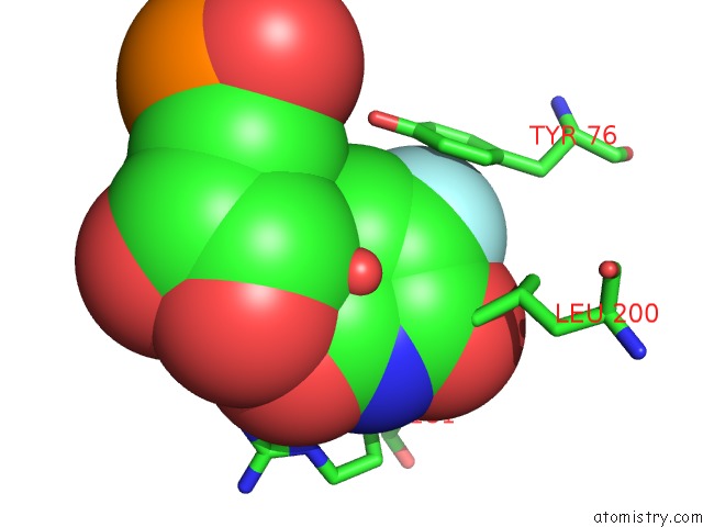
Mono view
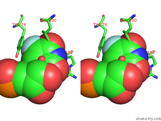
Stereo pair view
Mono view
Stereo pair view
A full contact list of Fluorine with other atoms in the F binding
site number 1 of Coupling of Active Site Motions and Rna Binding within 5.0Å range:
|
Reference:
C.Hoang,
C.S.Hamilton,
E.G.Mueller,
A.R.Ferre-D'amare.
Precursor Complex Structure of Pseudouridine Synthase Trub Suggests Coupling of Active Site Perturbations to An Rna-Sequestering Peripheral Protein Domain Protein Sci. V. 14 2201 2005.
ISSN: ISSN 0961-8368
PubMed: 15987897
DOI: 10.1110/PS.051493605
Page generated: Wed Jul 31 13:39:03 2024
ISSN: ISSN 0961-8368
PubMed: 15987897
DOI: 10.1110/PS.051493605
Last articles
Zn in 9MJ5Zn in 9HNW
Zn in 9G0L
Zn in 9FNE
Zn in 9DZN
Zn in 9E0I
Zn in 9D32
Zn in 9DAK
Zn in 8ZXC
Zn in 8ZUF