Fluorine »
PDB 2a4z-2baq »
2a7q »
Fluorine in PDB 2a7q: Crystal Structure of Human Dck Complexed with Clofarabine and Adp
Enzymatic activity of Crystal Structure of Human Dck Complexed with Clofarabine and Adp
All present enzymatic activity of Crystal Structure of Human Dck Complexed with Clofarabine and Adp:
2.7.1.74;
2.7.1.74;
Protein crystallography data
The structure of Crystal Structure of Human Dck Complexed with Clofarabine and Adp, PDB code: 2a7q
was solved by
Y.Zhang,
J.A.Secrist Iii,
S.E.Ealick,
with X-Ray Crystallography technique. A brief refinement statistics is given in the table below:
| Resolution Low / High (Å) | 48.49 / 2.55 |
| Space group | P 43 21 2 |
| Cell size a, b, c (Å), α, β, γ (°) | 80.145, 80.145, 93.669, 90.00, 90.00, 90.00 |
| R / Rfree (%) | 21.3 / 24.6 |
Other elements in 2a7q:
The structure of Crystal Structure of Human Dck Complexed with Clofarabine and Adp also contains other interesting chemical elements:
| Magnesium | (Mg) | 1 atom |
| Chlorine | (Cl) | 1 atom |
Fluorine Binding Sites:
The binding sites of Fluorine atom in the Crystal Structure of Human Dck Complexed with Clofarabine and Adp
(pdb code 2a7q). This binding sites where shown within
5.0 Angstroms radius around Fluorine atom.
In total only one binding site of Fluorine was determined in the Crystal Structure of Human Dck Complexed with Clofarabine and Adp, PDB code: 2a7q:
In total only one binding site of Fluorine was determined in the Crystal Structure of Human Dck Complexed with Clofarabine and Adp, PDB code: 2a7q:
Fluorine binding site 1 out of 1 in 2a7q
Go back to
Fluorine binding site 1 out
of 1 in the Crystal Structure of Human Dck Complexed with Clofarabine and Adp
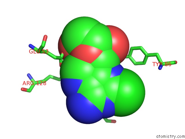
Mono view
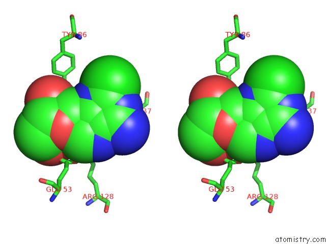
Stereo pair view
Mono view
Stereo pair view
A full contact list of Fluorine with other atoms in the F binding
site number 1 of Crystal Structure of Human Dck Complexed with Clofarabine and Adp within 5.0Å range:
|
Reference:
Y.Zhang,
J.A.Secrist,
S.E.Ealick.
The Structure of Human Deoxycytidine Kinase in Complex with Clofarabine Reveals Key Interactions For Prodrug Activation. Acta Crystallogr.,Sect.D V. 62 133 2006.
ISSN: ISSN 0907-4449
PubMed: 16421443
DOI: 10.1107/S0907444905034293
Page generated: Wed Jul 31 13:43:21 2024
ISSN: ISSN 0907-4449
PubMed: 16421443
DOI: 10.1107/S0907444905034293
Last articles
Zn in 9J0NZn in 9J0O
Zn in 9J0P
Zn in 9FJX
Zn in 9EKB
Zn in 9C0F
Zn in 9CAH
Zn in 9CH0
Zn in 9CH3
Zn in 9CH1