Fluorine »
PDB 2a4z-2baq »
2as4 »
Fluorine in PDB 2as4: Cytochrome C Peroxidase in Complex with 3-Fluorocatechol
Enzymatic activity of Cytochrome C Peroxidase in Complex with 3-Fluorocatechol
All present enzymatic activity of Cytochrome C Peroxidase in Complex with 3-Fluorocatechol:
1.11.1.5;
1.11.1.5;
Protein crystallography data
The structure of Cytochrome C Peroxidase in Complex with 3-Fluorocatechol, PDB code: 2as4
was solved by
R.Brenk,
S.W.Vetter,
S.E.Boyce,
D.B.Goodin,
B.K.Shoichet,
with X-Ray Crystallography technique. A brief refinement statistics is given in the table below:
| Resolution Low / High (Å) | 10.00 / 1.30 |
| Space group | P 21 21 21 |
| Cell size a, b, c (Å), α, β, γ (°) | 51.240, 75.490, 107.040, 90.00, 90.00, 90.00 |
| R / Rfree (%) | n/a / 17.5 |
Other elements in 2as4:
The structure of Cytochrome C Peroxidase in Complex with 3-Fluorocatechol also contains other interesting chemical elements:
| Potassium | (K) | 1 atom |
| Iron | (Fe) | 1 atom |
Fluorine Binding Sites:
The binding sites of Fluorine atom in the Cytochrome C Peroxidase in Complex with 3-Fluorocatechol
(pdb code 2as4). This binding sites where shown within
5.0 Angstroms radius around Fluorine atom.
In total 2 binding sites of Fluorine where determined in the Cytochrome C Peroxidase in Complex with 3-Fluorocatechol, PDB code: 2as4:
Jump to Fluorine binding site number: 1; 2;
In total 2 binding sites of Fluorine where determined in the Cytochrome C Peroxidase in Complex with 3-Fluorocatechol, PDB code: 2as4:
Jump to Fluorine binding site number: 1; 2;
Fluorine binding site 1 out of 2 in 2as4
Go back to
Fluorine binding site 1 out
of 2 in the Cytochrome C Peroxidase in Complex with 3-Fluorocatechol
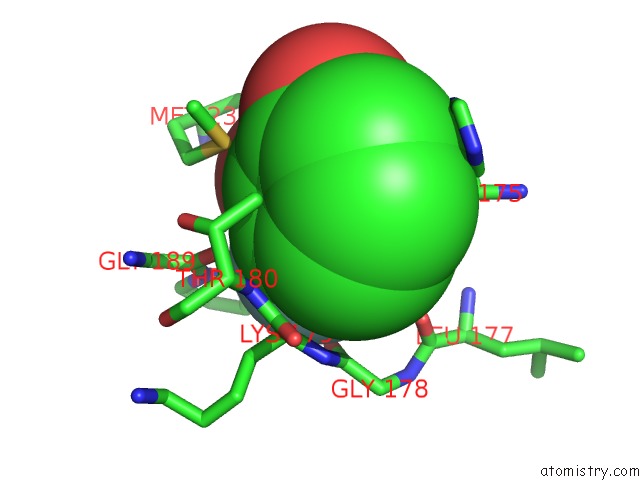
Mono view
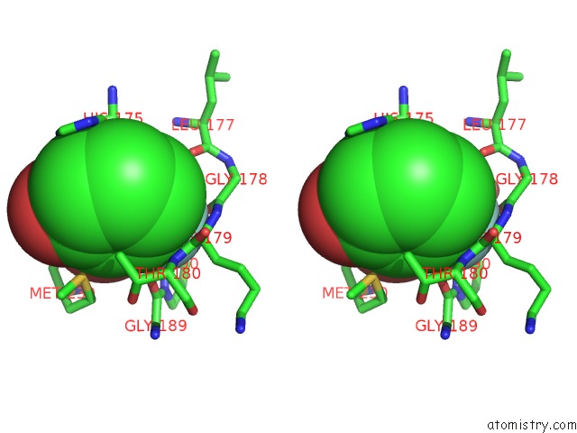
Stereo pair view
Mono view
Stereo pair view
A full contact list of Fluorine with other atoms in the F binding
site number 1 of Cytochrome C Peroxidase in Complex with 3-Fluorocatechol within 5.0Å range:
|
Fluorine binding site 2 out of 2 in 2as4
Go back to
Fluorine binding site 2 out
of 2 in the Cytochrome C Peroxidase in Complex with 3-Fluorocatechol
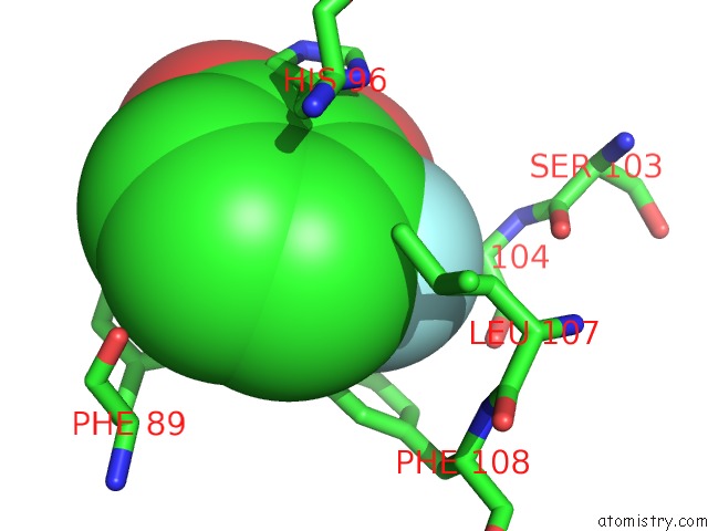
Mono view
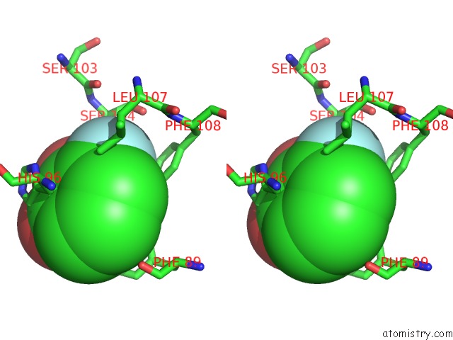
Stereo pair view
Mono view
Stereo pair view
A full contact list of Fluorine with other atoms in the F binding
site number 2 of Cytochrome C Peroxidase in Complex with 3-Fluorocatechol within 5.0Å range:
|
Reference:
R.Brenk,
S.W.Vetter,
S.E.Boyce,
D.B.Goodin,
B.K.Shoichet.
Probing Molecular Docking in A Charged Model Binding Site. J.Mol.Biol. V. 357 1449 2006.
ISSN: ISSN 0022-2836
PubMed: 16490206
DOI: 10.1016/J.JMB.2006.01.034
Page generated: Wed Jul 31 13:45:25 2024
ISSN: ISSN 0022-2836
PubMed: 16490206
DOI: 10.1016/J.JMB.2006.01.034
Last articles
Zn in 9MJ5Zn in 9HNW
Zn in 9G0L
Zn in 9FNE
Zn in 9DZN
Zn in 9E0I
Zn in 9D32
Zn in 9DAK
Zn in 8ZXC
Zn in 8ZUF