Fluorine »
PDB 2a4z-2baq »
2ayl »
Fluorine in PDB 2ayl: 2.0 Angstrom Crystal Structure of Manganese Protoporphyrin IX- Reconstituted Ovine Prostaglandin H2 Synthase-1 Complexed with Flurbiprofen
Enzymatic activity of 2.0 Angstrom Crystal Structure of Manganese Protoporphyrin IX- Reconstituted Ovine Prostaglandin H2 Synthase-1 Complexed with Flurbiprofen
All present enzymatic activity of 2.0 Angstrom Crystal Structure of Manganese Protoporphyrin IX- Reconstituted Ovine Prostaglandin H2 Synthase-1 Complexed with Flurbiprofen:
1.14.99.1;
1.14.99.1;
Protein crystallography data
The structure of 2.0 Angstrom Crystal Structure of Manganese Protoporphyrin IX- Reconstituted Ovine Prostaglandin H2 Synthase-1 Complexed with Flurbiprofen, PDB code: 2ayl
was solved by
K.Gupta,
B.S.Selinsky,
P.J.Loll,
with X-Ray Crystallography technique. A brief refinement statistics is given in the table below:
| Resolution Low / High (Å) | 43.33 / 2.00 |
| Space group | I 2 2 2 |
| Cell size a, b, c (Å), α, β, γ (°) | 98.931, 206.550, 221.553, 90.00, 90.00, 90.00 |
| R / Rfree (%) | 21.8 / 23.7 |
Other elements in 2ayl:
The structure of 2.0 Angstrom Crystal Structure of Manganese Protoporphyrin IX- Reconstituted Ovine Prostaglandin H2 Synthase-1 Complexed with Flurbiprofen also contains other interesting chemical elements:
| Manganese | (Mn) | 2 atoms |
Fluorine Binding Sites:
The binding sites of Fluorine atom in the 2.0 Angstrom Crystal Structure of Manganese Protoporphyrin IX- Reconstituted Ovine Prostaglandin H2 Synthase-1 Complexed with Flurbiprofen
(pdb code 2ayl). This binding sites where shown within
5.0 Angstroms radius around Fluorine atom.
In total 4 binding sites of Fluorine where determined in the 2.0 Angstrom Crystal Structure of Manganese Protoporphyrin IX- Reconstituted Ovine Prostaglandin H2 Synthase-1 Complexed with Flurbiprofen, PDB code: 2ayl:
Jump to Fluorine binding site number: 1; 2; 3; 4;
In total 4 binding sites of Fluorine where determined in the 2.0 Angstrom Crystal Structure of Manganese Protoporphyrin IX- Reconstituted Ovine Prostaglandin H2 Synthase-1 Complexed with Flurbiprofen, PDB code: 2ayl:
Jump to Fluorine binding site number: 1; 2; 3; 4;
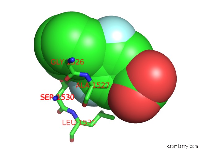
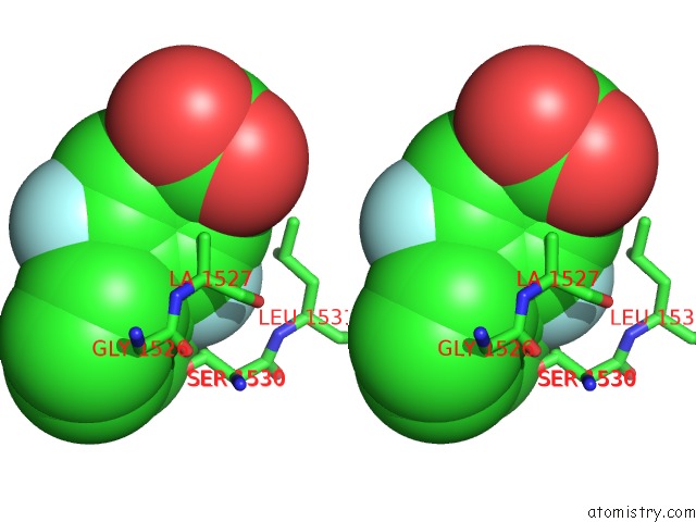
Fluorine binding site 1 out of 4 in 2ayl
Go back to
Fluorine binding site 1 out
of 4 in the 2.0 Angstrom Crystal Structure of Manganese Protoporphyrin IX- Reconstituted Ovine Prostaglandin H2 Synthase-1 Complexed with Flurbiprofen
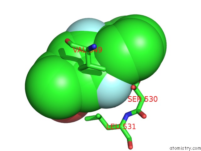
Mono view
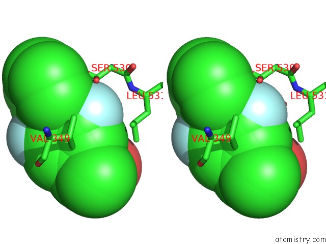
Stereo pair view
Mono view
Stereo pair view
A full contact list of Fluorine with other atoms in the F binding
site number 1 of 2.0 Angstrom Crystal Structure of Manganese Protoporphyrin IX- Reconstituted Ovine Prostaglandin H2 Synthase-1 Complexed with Flurbiprofen within 5.0Å range:
|
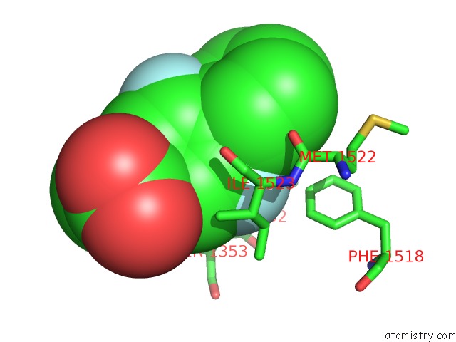
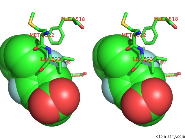
Fluorine binding site 2 out of 4 in 2ayl
Go back to
Fluorine binding site 2 out
of 4 in the 2.0 Angstrom Crystal Structure of Manganese Protoporphyrin IX- Reconstituted Ovine Prostaglandin H2 Synthase-1 Complexed with Flurbiprofen
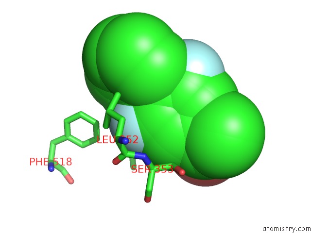
Mono view
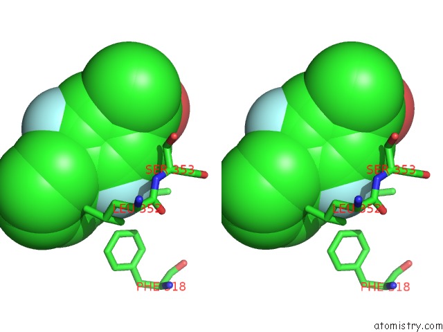
Stereo pair view
Mono view
Stereo pair view
A full contact list of Fluorine with other atoms in the F binding
site number 2 of 2.0 Angstrom Crystal Structure of Manganese Protoporphyrin IX- Reconstituted Ovine Prostaglandin H2 Synthase-1 Complexed with Flurbiprofen within 5.0Å range:
|
Fluorine binding site 3 out of 4 in 2ayl
Go back to
Fluorine binding site 3 out
of 4 in the 2.0 Angstrom Crystal Structure of Manganese Protoporphyrin IX- Reconstituted Ovine Prostaglandin H2 Synthase-1 Complexed with Flurbiprofen
Mono view
Stereo pair view
Mono view
Stereo pair view
A full contact list of Fluorine with other atoms in the F binding
site number 3 of 2.0 Angstrom Crystal Structure of Manganese Protoporphyrin IX- Reconstituted Ovine Prostaglandin H2 Synthase-1 Complexed with Flurbiprofen within 5.0Å range:
|
Fluorine binding site 4 out of 4 in 2ayl
Go back to
Fluorine binding site 4 out
of 4 in the 2.0 Angstrom Crystal Structure of Manganese Protoporphyrin IX- Reconstituted Ovine Prostaglandin H2 Synthase-1 Complexed with Flurbiprofen
Mono view
Stereo pair view
Mono view
Stereo pair view
A full contact list of Fluorine with other atoms in the F binding
site number 4 of 2.0 Angstrom Crystal Structure of Manganese Protoporphyrin IX- Reconstituted Ovine Prostaglandin H2 Synthase-1 Complexed with Flurbiprofen within 5.0Å range:
|
Reference:
K.Gupta,
B.S.Selinsky,
P.J.Loll.
2.0 Angstroms Structure of Prostaglandin H2 Synthase-1 Reconstituted with A Manganese Porphyrin Cofactor. Acta Crystallogr.,Sect.D V. 62 151 2006.
ISSN: ISSN 0907-4449
PubMed: 16421446
DOI: 10.1107/S0907444905036309
Page generated: Wed Jul 31 13:47:37 2024
ISSN: ISSN 0907-4449
PubMed: 16421446
DOI: 10.1107/S0907444905036309
Last articles
Zn in 9JYWZn in 9IR4
Zn in 9IR3
Zn in 9GMX
Zn in 9GMW
Zn in 9JEJ
Zn in 9ERF
Zn in 9ERE
Zn in 9EGV
Zn in 9EGW